Difference between revisions of "Steatocystoma"
Jump to navigation
Jump to search
(split out) |
|||
| Line 1: | Line 1: | ||
{{ Infobox diagnosis | |||
| Name = {{PAGENAME}} | |||
| Image = Steatocystoma - very high mag.jpg | |||
| Width = | |||
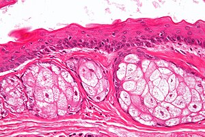
| Caption = Steatocystoma. [[H&E stain]]. | |||
| Synonyms = | |||
| Micro = cyst lined by squamous epithelium with a corrugated eosinophilic lining, no granular cell layer | |||
| Subtypes = | |||
| LMDDx = | |||
| Stains = | |||
| IHC = | |||
| EM = | |||
| Molecular = | |||
| IF = | |||
| Gross = | |||
| Grossing = | |||
| Site = [[skin]] - see ''[[dermal cysts]]'' | |||
| Assdx = | |||
| Syndromes = steatocystoma multiplex | |||
| Clinicalhx = | |||
| Signs = | |||
| Symptoms = | |||
| Prevalence = rare | |||
| Bloodwork = | |||
| Rads = | |||
| Endoscopy = | |||
| Prognosis = benign | |||
| Other = | |||
| ClinDDx = | |||
| Tx = | |||
}} | |||
'''Steatocystoma''' is a rare benign [[dermal cyst]]. | '''Steatocystoma''' is a rare benign [[dermal cyst]]. | ||
| Line 21: | Line 52: | ||
Image:Steatocystoma_-_low_mag.jpg | Steatocystoma - low mag. (WC/Nephron) | Image:Steatocystoma_-_low_mag.jpg | Steatocystoma - low mag. (WC/Nephron) | ||
Image:Steatocystoma_-_intermed_mag.jpg | Steatocystoma - intermed. mag. (WC/Nephron) | Image:Steatocystoma_-_intermed_mag.jpg | Steatocystoma - intermed. mag. (WC/Nephron) | ||
Image:Steatocystoma_-_high_mag.jpg | Steatocystoma - high mag. (WC/Nephron) | |||
</gallery> | </gallery> | ||
www: | www: | ||
Revision as of 22:51, 16 February 2014
| Steatocystoma | |
|---|---|
| Diagnosis in short | |
 Steatocystoma. H&E stain. | |
|
| |
| LM | cyst lined by squamous epithelium with a corrugated eosinophilic lining, no granular cell layer |
| Site | skin - see dermal cysts |
|
| |
| Syndromes | steatocystoma multiplex |
|
| |
| Prevalence | rare |
| Prognosis | benign |
Steatocystoma is a rare benign dermal cyst.
General
- Benign.
- Typically adults.
- Usually on the trunk.
- May be genetic; known as steatocystoma multiplex.[1]
- Classically autosomal dominant.[2]
Microscopic
Features:[3]
- Cyst lined by squamous epithelium with:
- Corrugated eosinophilic lining - key feature.
- Similar appearance to compact keratin (hyperkeratosis).
- Described as a hyaline cuticle.[4]
- No granular cell layer.
- Corrugated eosinophilic lining - key feature.
Images
www:
See also
References
- ↑ Online 'Mendelian Inheritance in Man' (OMIM) 184500
- ↑ URL: http://path.upmc.edu/cases/case674/dx.html. Accessed on: 29 January 2012.
- ↑ Busam, Klaus J. (2009). Dermatopathology: A Volume in the Foundations in Diagnostic Pathology Series (1st ed.). Saunders. pp. 312. ISBN 978-0443066542.
- ↑ URL: http://path.upmc.edu/cases/case674/dx.html. Accessed on: 29 January 2012.
- ↑ URL: http://path.upmc.edu/cases/case674.html. Accessed on: 29 January 2012.


