Difference between revisions of "Actinic keratosis"
| Line 83: | Line 83: | ||
*Pigmented actinic keratosis. | *Pigmented actinic keratosis. | ||
**DDx: [[lentigo maligna]]. | **DDx: [[lentigo maligna]]. | ||
*Lichenoid actinic keratosis. | *Lichenoid actinic keratosis - band of inflammatory cells at the dermal-epidermal junction. | ||
**DDx: [[lichen planus]]. | |||
==Sign out== | ==Sign out== | ||
Revision as of 21:12, 29 October 2013
| Actinic keratosis | |
|---|---|
| Diagnosis in short | |
 | |
|
| |
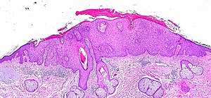
| LM | basal epidermal atypia - key feature, palisading of basal cells, solar elastosis +/-parakeratosis |
| Subtypes | bowenoid actinic keratosis, hypertrophic actinic keratosis, acantholytic actinic keratosis, proliferative actinic keratosis, pigmented actinic keratosis, lichenoid actinic keratosis |
| LM DDx | squamous cell carcinoma of the skin, Bowen's disease |
| IHC | CK34betaE12 +ve |
| Site | skin |
|
| |
| Associated Dx | squamous cell carcinoma of the skin, Bowen's disease |
| Signs | sandpaper-like texture |
| Prognosis | good |
| Clin. DDx | squamous cell carcinoma, melanoma (for pigmented AK) |
Actinic keratosis, abbreviated AK, is a common skin lesion. It is also known as solar keratosis.[1][2]
General
- Considered a precursor of squamous cell carcinoma of the skin.[2]
Risk factors:[3]
- Sun exposure.
- Immune suppression (e.g. organ transplant recipients).
Gross
Features:
- Yellow-brown scaly, patches.
- Sandpaper sensation - on touching.
- May be pigmented.[4]
Microscopic
Features:[5]
- Epidermal nuclear atypia:
- Variation is size, shape and staining - must involve basilar layer.
- Nuclear enlargement - key feature.
- Hyperchromasia.
- Variation is size, shape and staining - must involve basilar layer.
- Abnormal epidermal architecture:
- Palisading.[citation needed]
- +/-Parakeratosis - may alternate with orthokeratosis, esp. early lesions.[6]
- +/-Irregular acanthosis.
Note:
- May be full thickness - known as bowenoid actinic keratosis.[7]
DDx:
- Actinic cheilitis - the same lesion on the lip.[8]
- Bowen's disease - full thickness involvement - with involvement of adnexal epithelium and follicular epithelium.[7]
- Paget disease of the breast.
- Squamous cell carcinoma of the skin.
- Lentigo maligna (melanoma in situ on sun damaged skin) - esp. for pigmented AK.[6]
- One should see benign melanocytes within an AK... otherwise one should do stains as the melanocytes may be mimicking keratinocytes.
- Seborrheic keratosis - should not have basilar nuclear atypia.
- Lichenoid keratosis (esp. for lichenoid actinic keratosis) - has a band of inflammatory cells in the superficial dermis.
Images
Histologic subtypes of actinic keratosis
Like most common things, there are several variants:[6]
- Hypertrophic actinic keratosis.
- Increased thickness of: (1) epidermis and, (2) stratum corneum.[9]
- Acantholytic actinic keratosis.
- Proliferative actinic keratosis - downward finger-like projections of the epidermis.
- Pigmented actinic keratosis.
- DDx: lentigo maligna.
- Lichenoid actinic keratosis - band of inflammatory cells at the dermal-epidermal junction.
- DDx: lichen planus.
Sign out
SKIN LESION, RIGHT THIGH, BIOPSY:
- HYPERTROPHIC ACTINIC KERATOSIS.
- SOLAR ELASTOSIS.
- NEGATIVE FOR MALIGNANCY.
Bowenoid
SKIN LESION, RIGHT DISTAL FOREARM, BIOPSY:
- BOWENOID ACTINIC KERATOSIS, COMPLETELY EXCISED IN PLANE OF SECTION.
- EXTENSIVE SOLAR ELASTOSIS.
- NEGATIVE FOR MALIGNANCY.
Micro
General
The sections show skin with basilar epidermal nuclear enlargement and hyperchromasia with irregular nuclear palisading. Hyperkeratosis and parakeratosis is present. A granular layer is present. The dermal-epidermal interface is sharply-demarcated. There is focal inflammation at the interface. Extensive solar elastosis is present.
There is no clefting of the lesion from the surrounding stroma. The surrounding stroma does not have a myxoid quality.
Bowenoid
The sections show skin with epidermal nuclear enlargement and hyperchromasia with irregular nuclear palisading. The lesion involves the full thickness of the epidermis; however, it spares adnexal and follicular epithelium. Proliferative activity is present. Hyperkeratosis and parakeratosis is present. A granular layer is present. The dermal-epidermal interface is sharply-demarcated. There is focal inflammation at the interface. Extensive solar elastosis is present.
There is no clefting of the lesion from the surrounding stroma. Intercellular bridges are identified.
See also
References
- ↑ Weinberg, JM. (Jan 2006). "Topical therapy for actinic keratoses: current and evolving therapies.". Rev Recent Clin Trials 1 (1): 53-60. PMID 18393780.
- ↑ 2.0 2.1 Roewert-Huber, J.; Stockfleth, E.; Kerl, H. (Dec 2007). "Pathology and pathobiology of actinic (solar) keratosis - an update.". Br J Dermatol 157 Suppl 2: 18-20. doi:10.1111/j.1365-2133.2007.08267.x. PMID 18067626.
- ↑ Kumar, Vinay; Abbas, Abul K.; Fausto, Nelson; Aster, Jon (2009). Robbins and Cotran pathologic basis of disease (8th ed.). Elsevier Saunders. pp. 1180. ISBN 978-1416031215.
- ↑ URL: http://www.dermoscopy-ids.org/index.php/newest-case/79-lentigo-maligna-vs-pigmented-actinic-keratosis. Accessed on: 23 August 2012.
- ↑ URL: http://emedicine.medscape.com/article/1099775-workup#a0723. Accessed on: 1 September 2011.
- ↑ 6.0 6.1 6.2 Busam, Klaus J. (2009). Dermatopathology: A Volume in the Foundations in Diagnostic Pathology Series (1st ed.). Saunders. pp. 353. ISBN 978-0443066542.
- ↑ 7.0 7.1 Bagazgoitia, L.; Cuevas, J.; Juarranz, A. (Feb 2010). "Expression of p53 and p16 in actinic keratosis, bowenoid actinic keratosis and Bowen's disease.". J Eur Acad Dermatol Venereol 24 (2): 228-30. doi:10.1111/j.1468-3083.2009.03337.x. PMID 19515076.
- ↑ Picascia, DD.; Robinson, JK. (Aug 1987). "Actinic cheilitis: a review of the etiology, differential diagnosis, and treatment.". J Am Acad Dermatol 17 (2 Pt 1): 255-64. PMID 3305604.
- ↑ Busam, Klaus J. (2009). Dermatopathology: A Volume in the Foundations in Diagnostic Pathology Series (1st ed.). Saunders. pp. 352. ISBN 978-0443066542.


