Difference between revisions of "Oral pathology"
Jump to navigation
Jump to search
(→Melanotic macule: +SO) |
|||
| Line 115: | Line 115: | ||
===General=== | ===General=== | ||
*Benign. | *Benign. | ||
*Clinically apparent lesion. | |||
===Gross=== | ===Gross=== | ||
*Flat pigmented lesion less than 10 mm in size ([[macule]]). | *Flat pigmented lesion less than 10 mm in size ([[macule]]). | ||
*Usually solitary.<ref name=pmid289929>{{Cite journal | last1 = Buchner | first1 = A. | last2 = Hansen | first2 = LS. | title = Melanotic macule of the oral mucosa. A clinicopathologic study of 105 cases. | journal = Oral Surg Oral Med Oral Pathol | volume = 48 | issue = 3 | pages = 244-9 | month = Sep | year = 1979 | doi = | PMID = 289929 }}</ref> | |||
Image: | Image: | ||
| Line 123: | Line 125: | ||
===Microscopic=== | ===Microscopic=== | ||
Features: | Features - either or both of the following:<ref name=pmid289929/> | ||
#Pigmented basal cell layer. | |||
#Pigment incontinence - (dermal) macrophages with pigment (melanin). | |||
DDx: | |||
*Focal melanosis - ''not'' clinically apparent. | |||
Image: | Image: | ||
*[http://ocw.tufts.edu/Content/51/lecturenotes/551831/552086 Melanotic macule - labia (tufts.edu)]. | *[http://ocw.tufts.edu/Content/51/lecturenotes/551831/552086 Melanotic macule - labia (tufts.edu)]. | ||
===Sign out=== | |||
<pre> | |||
LESION, BUCCAL MUCOSA (LEFT), BIOPSY: | |||
- MELANOTIC MACUOLE. | |||
- NEGATIVE FOR MALIGNANCY. | |||
</pre> | |||
==Smoker's melanosis== | ==Smoker's melanosis== | ||
Revision as of 09:43, 30 July 2013
Oral pathology is a domain of dentistry. In the context of anatomical pathology, it can be lumped with head and neck pathology. Oral lesions redirects here.
Odontogenic tumours and cysts
Main article: Odontogenic tumours and cysts
Oral infections
Oral candidiasis
General
- Due to candida - a fungus.
- May be associated with immunodeficiency, e.g. AIDS, organ transplant/immunosuppression.
Forms:[1]
- Pseudomembranous (thrush).
- Erythematous.
- Hyperplastic.
Microscopic
- See candidiasis.
Hairy leukoplakia
General
Features:[1]
Gross
- White confluent patches (icing sugar) - usu. tongue.
DDx:
- See leukoplakia.
Images:
Microscopic
Features:[4]
- Hyperkeratosis (thicker stratum corneum).[5]
- Acanthosis (thicker stratum spinosum).[6]
- "Balloon cells" in upper stratum spinosum - perinuclear clearing.
Oral condyloma
General
- Benign.
- Sexually transmitted.[7]
- Typically seen in young adults.
Gross
- Polypoid projection with a broad base.
- Usually palate or labia.[7]
Microscopic
Features:
- Broad papillary projections with rounded contours.
- No hyperkeratosis.
DDx:[7]
- Squamous papilloma - thinner papillary projections, often branch.
- Verruca vulgaris - church spire-like projections, hyperkeratosis and parakeratosis.
- Squamous cell carcinoma.
Image:
Sign out
LESION, PALATE, EXCISION:
- ORAL CONDYLOMA.
Oral neoplasms
Peripheral fibroma
- AKA focal fibrous hyperplasia, AKA peripheral ossifying fibroma, AKA fibroid epulis (old term), AKA fibroepithelial polyp.[9]
- AKA oral fibroma.[10][11]
General
- Most common oral cavity tumour.[11]
- Female predominance (female:male = 2:1), usually 30-50 years old.[11]
- Multiple oral fibromas may be seen in Cowden disease.[12][13]
- Histologically similar to fibrous papule.[14]
Microscopic
Features:[14]
- Fibrous stroma - key feature.
- "Very pink" at low power.
- +/-Collagen bundles, may be prominent.
- Prominent (dilated) vessels.
- Overlying (squamous) mucosa benign (flat).
- +/-Hyperkeratosis +/-focal ulceration.[11]
Sign out
TONGUE LESION, BIOPSY:
- FIBROMA.
Pigmented lesions of the oral cavity
A brief DDx of pigmented lesions:[15]
- Diffuse & bilateral:
- Peutz-Jeghers syndrome.
- Addison's disease.
- Drug-induced - typically OCP or tetracycline, usu. has an irregular distribution.[16]
- Smoker's melanosis.
- Focal:
- Vascular lesions.
- Amalgam tattoo.
- Melanocytic lesions.
- Melanotic macule.
- Blue nevus.
- Malignant melanoma - classically hard palate.[16]
Melanotic macule
General
- Benign.
- Clinically apparent lesion.
Gross
Image:
Microscopic
Features - either or both of the following:[17]
- Pigmented basal cell layer.
- Pigment incontinence - (dermal) macrophages with pigment (melanin).
DDx:
- Focal melanosis - not clinically apparent.
Image:
Sign out
LESION, BUCCAL MUCOSA (LEFT), BIOPSY:
- MELANOTIC MACUOLE.
- NEGATIVE FOR MALIGNANCY.
Smoker's melanosis
General
- Benign.
- Seen in ~20% of smokers.[15]
- Presence of find (smoking) dose-dependent, i.e. longer heavier smokers are more likely to have it.
Gross
- Typically labial gingvia or buccal mucosa.[15]
Microscopic
Features:
- Basal melanosis.
- +/-Melanin incontinence.
Image:
Intramucosal melanocytic nevus
- Abbreviated IMN.
- AKA intramucosal melanocytic nevus.
General
- Most common oral nevus.[18]
- Second most common is the blue nevus.
- Essentially an intradermal melanocytic nevus.
Microscopic
Features:
- Symmetrical lesion.
- "Matures" with depth
- Less cellular with depth
- Less nuclear atypia with depth.
- Smaller cells with depth.
- Smaller nests with depth.
- Rare mitoses (superficial).
- No deep mitoses.
- No destruction of surrounding structures.
- No nucleoli.
Sign out
PALATE LESION, PUNCH BIOPSY:
- INTRAMUCOSAL MELANOCYTIC NEVUS.
Amalgam tattoo
General
- Benign and common.
- Material from a dental filling.[19]
- May be confused with a melanocytic lesion.
Gross
- Pigmented lesion.
Image:
Microscopic
Features:[20]
- Fine powdery black material in the subepithelial tissue - key feature.
- May be clumped.
- Found between collagen fibres.
- +/-Foreign body-type giant cells - uncommon.
DDx:
Images
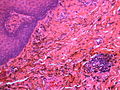
Amalgam tattoo. (WC/Bernhard138)
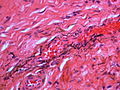
Amalgam tattoo. (WC/Bernhard138)
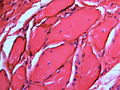
Amalgam tattoo. (WC/Bernhard138)


www:
Sign out
MOUTH, BIOPSY:
- AMALGAM TATTOO.
- SQUAMOUS MUCOSA WITH PARAKERATOSIS.
- SUBEPITHELIAL CALCIFICATIONS.
- NEGATIVE FOR MALIGNANCY.
See also
References
- ↑ 1.0 1.1 Cotran, Ramzi S.; Kumar, Vinay; Fausto, Nelson; Nelso Fausto; Robbins, Stanley L.; Abbas, Abul K. (2005). Robbins and Cotran pathologic basis of disease (7th ed.). St. Louis, Mo: Elsevier Saunders. pp. 777. ISBN 0-7216-0187-1.
- ↑ Kanitakis, J.; Zambruno, G.; Marchand, C.; Perret-Liaudet, P.; Hermier, C.; Thivolet, J. (1990). "[Oral hairy leukoplakia in AIDS. Histologic and ultrastructural study of 8 cases].". Ann Dermatol Venereol 117 (5): 345-53. PMID 2169222.
- ↑ Itin, PH.; Lautenschlager, S. (1997). "Viral lesions of the mouth in HIV-infected patients.". Dermatology 194 (1): 1-7. PMID 9031782.
- ↑ URL: http://www.pathologyoutlines.com/oralcavity.html#hairyleukoplakia.
- ↑ URL: http://www.emedicine.com/asp/dictionary.asp?keyword=hyperkeratosis.
- ↑ URL: http://www.emedicine.com/asp/dictionary.asp?keyword=acanthosis.
- ↑ 7.0 7.1 7.2 Thompson, Lester D. R. (2006). Head and Neck Pathology: A Volume in Foundations in Diagnostic Pathology Series (1st ed.). Churchill Livingstone. pp. 426. ISBN 978-0443069604.
- ↑ Reis, HL.; Ferreira, DC.; Forattini, AG.; Souza, PG.; Curvelo, JA.; Passos, MR. (2010). "Genital and oral human papillomavirus infection in a patient from the group of women who have sex with women.". Clinics (Sao Paulo) 65 (12): 1383-5. PMC 3020353. PMID 21340231. https://www.ncbi.nlm.nih.gov/pmc/articles/PMC3020353/.
- ↑ Mills, Stacey E; Carter, Darryl; Greenson, Joel K; Reuter, Victor E; Stoler, Mark H (2009). Sternberg's Diagnostic Surgical Pathology (5th ed.). Lippincott Williams & Wilkins. pp. 775. ISBN 978-0781779425.
- ↑ URL: http://emedicine.medscape.com/article/1080948-overview#aw2aab6b3. Accessed on: 20 August 2012.
- ↑ 11.0 11.1 11.2 11.3 Thompson, Lester D. R. (2006). Head and Neck Pathology: A Volume in Foundations in Diagnostic Pathology Series (1st ed.). Churchill Livingstone. pp. 240. ISBN 978-0443069604.
- ↑ Segura Saint-Gerons, R.; Ceballos Salobreña, A.; Toro Rojas, M.; Gándara Rey, JM. (Aug 2006). "Oral manifestations of Cowden's disease. Presentation of a clinical case.". Med Oral Patol Oral Cir Bucal 11 (5): E421-4. PMID 16878060.
- ↑ Oliveira, MA.; Medina, JB.; Xavier, FC.; Magalhães, M.; Ortega, KL. (2010). "Cowden syndrome.". Dermatol Online J 16 (1): 7. PMID 20137749.
- ↑ 14.0 14.1 Fernandez-Flores, A. (Jul 2010). "Solitary oral fibromas of the tongue show similar morphologic features to fibrous papule of the face: a study of 31 cases.". Am J Dermatopathol 32 (5): 442-7. doi:10.1097/DAD.0b013e3181c47142. PMID 20421776.
- ↑ 15.0 15.1 15.2 Kauzman, A.; Pavone, M.; Blanas, N.; Bradley, G. (Nov 2004). "Pigmented lesions of the oral cavity: review, differential diagnosis, and case presentations.". J Can Dent Assoc 70 (10): 682-3. PMID 15530266.
- ↑ 16.0 16.1 Beck-Mannagetta, J.; Hutarew, G. (Sep 2012). "[Pigmented lesions of the oral mucosa].". Hautarzt 63 (9): 704-9. doi:10.1007/s00105-012-2351-x. PMID 22956033.
- ↑ 17.0 17.1 Buchner, A.; Hansen, LS. (Sep 1979). "Melanotic macule of the oral mucosa. A clinicopathologic study of 105 cases.". Oral Surg Oral Med Oral Pathol 48 (3): 244-9. PMID 289929.
- ↑ URL: http://emedicine.medscape.com/article/1079272-overview. Accessed on: 10 December 2012.
- ↑ Ref NaNP|215
- ↑ Template:Ref NaNP