Difference between revisions of "Glomus tumour"
m (→Micro) |
(+infobox) |
||
| Line 1: | Line 1: | ||
{{ Infobox diagnosis | |||
| Name = {{PAGENAME}} | |||
| Image = Glomus_tumour_-_2_-_very_high_mag.jpg | |||
| Width = | |||
| Caption = Glomus tumour. [[H&E stain]]. | |||
| Micro = sheets of equally-spaced cells ("cookie cutter appearance"), polygonal cells with identifiable cellular borders, thin-walled blood vessels, moderate clear cytoplasm | |||
| Subtypes = | |||
| LMDDx = | |||
| Stains = | |||
| IHC = SMA +ve (100%), desmin usu. -ve, CD34 -ve, S-100 -ve | |||
| EM = | |||
| Molecular = | |||
| IF = | |||
| Gross = | |||
| Grossing = | |||
| Site = [[skin]]/soft tissue - classically subungal | |||
| Assdx = | |||
| Syndromes = | |||
| Clinicalhx = | |||
| Signs = | |||
| Symptoms = [[painful skin lesion]] | |||
| Prevalence = uncommon | |||
| Bloodwork = | |||
| Rads = | |||
| Endoscopy = | |||
| Prognosis = usu. good | |||
| Other = | |||
| ClinDDx = | |||
}} | |||
'''Glomus tumours''', also known as '''glomangioma''', are painful, perivascular tumour that are classically periungual. | '''Glomus tumours''', also known as '''glomangioma''', are painful, perivascular tumour that are classically periungual. | ||
| Line 33: | Line 62: | ||
*No significant nuclear atypia. | *No significant nuclear atypia. | ||
*The regular cell spacing is called "cookie cutter appearance". It looks like the cells were created with a cookie cutter; the spacing between cell is equal and they all look very similar. | *The regular cell spacing is called "cookie cutter appearance". It looks like the cells were created with a cookie cutter; the spacing between cell is equal and they all look very similar. | ||
*Should be perivascular - abut endothelial cells. | |||
Images | ===Images=== | ||
<gallery> | |||
Image:Glomus_tumour_-_intermed_mag.jpg | Glomus tumour - intermed. mag. (WC) | |||
Image:Glomus_tumour_-_2_-_very_high_mag.jpg | Glomus tumour - very high mag. (WC) | |||
</gallery> | |||
www: | |||
*[http://moon.ouhsc.edu/kfung/jty1/opaq/PathQuiz/Z0B003-PQ01-M.htm Glomus tumour (ouhsc.edu)]. | *[http://moon.ouhsc.edu/kfung/jty1/opaq/PathQuiz/Z0B003-PQ01-M.htm Glomus tumour (ouhsc.edu)]. | ||
Revision as of 02:05, 16 October 2013
| Glomus tumour | |
|---|---|
| Diagnosis in short | |
 Glomus tumour. H&E stain. | |
|
| |
| LM | sheets of equally-spaced cells ("cookie cutter appearance"), polygonal cells with identifiable cellular borders, thin-walled blood vessels, moderate clear cytoplasm |
| IHC | SMA +ve (100%), desmin usu. -ve, CD34 -ve, S-100 -ve |
| Site | skin/soft tissue - classically subungal |
|
| |
| Symptoms | painful skin lesion |
| Prevalence | uncommon |
| Prognosis | usu. good |
Glomus tumours, also known as glomangioma, are painful, perivascular tumour that are classically periungual.
It should not be confused with paraganglioma, which were once called glomus tumour.
This tumour is classified as a perivascular tumour (also pericytic tumour) which is a subset of soft tissue tumours.
General
- Tumour derived from smooth muscle cell.[1]
- Usually benign.
- Malignant variant exists - extremely rare.
Clinical:
Gross
Location:
- Classically subungual (under the nail).[2]
- Reported in almost very site imaginable.
- Most common GI site: stomach.[3]
Appearance (extradigital tumours):[2]
- Purplish papule.
Microscopic
Features:[4]
- Sheets of equally-spaced cells ("cookie cutter appearance") - key feature.
- Polygonal cells with identifiable cellular borders.
- Thin-walled blood vessels.
- May vaguely resemble antlers (staghorn vessels).
- Moderate clear cytoplasm.
Notes:
- No significant nuclear atypia.
- The regular cell spacing is called "cookie cutter appearance". It looks like the cells were created with a cookie cutter; the spacing between cell is equal and they all look very similar.
- Should be perivascular - abut endothelial cells.
Images
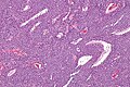
Glomus tumour - intermed. mag. (WC)
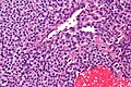
Glomus tumour - very high mag. (WC)

www:
IHC
Features:[5]
- SMA +ve ~ 100%.
- Desmin usu. -ve.
- CD34 -ve.
- Rarely +ve.
Others:
- S100 -ve.
Other diagnoses...
Why it is not a(n) ...[4]
- Angiosarcoma - has nuclear atypia.
- Dermatofibroma - spindle cell lesion.
- Capillary hemangioma - no epithelioid cells, more blood vessels.
Sign out
LESION, RIGHT RING FINGERNAIL, EXCISION:
- GLOMUS TUMOUR.

COMMENT:
The tumour cells are positive for SMA.
Micro
The sections show a tumour composed of polygonal cells with moderately distinct cellular borders and moderate cytoplasm. The tumour cell nuclei are round and have round nucleoli seen with the 10x objective. Thin-walled blood vessels with bland endothelial cells are found within the tumour. Tumour cells are identified immediately adjacent to the endothelial cells. No mitotic activity is apparent. Focal nuclear enlargement is present; however, no significant nuclear atypia is identified.
See also
- Painful skin lesions.
- Myopericytoma - the other perivascular (soft tissue) tumour.
References
- ↑ Gombos Z, Zhang PJ (September 2008). "Glomus tumor". Arch. Pathol. Lab. Med. 132 (9): 1448–52. doi:10.1043/1543-2165(2008)132[1448:GT]2.0.CO;2. PMID 18788860.
- ↑ 2.0 2.1 Lee, DW.; Yang, JH.; Chang, S.; Won, CH.; Lee, MW.; Choi, JH.; Moon, KC. (Dec 2011). "Clinical and pathological characteristics of extradigital and digital glomus tumours: a retrospective comparative study.". J Eur Acad Dermatol Venereol 25 (12): 1392-7. doi:10.1111/j.1468-3083.2011.03979.x. PMID 21371130.
- ↑ Miettinen, M.; Paal, E.; Lasota, J.; Sobin, LH. (Mar 2002). "Gastrointestinal glomus tumors: a clinicopathologic, immunohistochemical, and molecular genetic study of 32 cases.". Am J Surg Pathol 26 (3): 301-11. PMID 11859201.
- ↑ 4.0 4.1 URL: http://moon.ouhsc.edu/kfung/jty1/opaq/PathQuiz/Z0B003-PQ01-M.htm. Accessed on: 19 October 2010.
- ↑ Hatori M, Aiba S, Kato M, Kamiya N, Kokubun S (July 1997). "Expression of CD34 in glomus tumors". Tohoku J. Exp. Med. 182 (3): 241–7. PMID 9362106.