Difference between revisions of "Pineal gland"
Jump to navigation
Jump to search
(→Overview: w) |
(→Microscopic: +images) |
||
| Line 40: | Line 40: | ||
*Rosette = circular/flower-like arrangement of cells. | *Rosette = circular/flower-like arrangement of cells. | ||
Images | ====Images==== | ||
<gallery> | |||
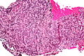
Image:Pineocytoma_-_intermed_mag.jpg | Pineocytoma - intermed. mag. (WC) | |||
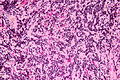
Image:Pineocytoma_-_high_mag.jpg | Pineocytoma - high mag. (WC) | |||
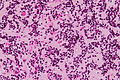
Image:Pineocytoma_-_very_high_mag.jpg | Pineocytoma - very high mag. (WC) | |||
</gallery> | |||
*www: | *www: | ||
**[http://path.upmc.edu/cases/case157.html Pineocytoma with neuronal differentiation (upmc.edu)]. | **[http://path.upmc.edu/cases/case157.html Pineocytoma with neuronal differentiation (upmc.edu)]. | ||
Revision as of 04:38, 18 November 2013
The pineal gland is thingy that is most noted for the fact that it calcifies with age.
Normal histology
- Cellular.
Overview
Tumours:[1]
- Primary pineal tumours ~15% of (pineal) tumours - benign to malignant:[2]
- Pineocytoma.
- Pineal parenchymal tumor of intermediate differentiation.
- Pineoblastoma.
- Germ cell tumours:
- Germinoma ~ 50% of (pineal) tumours.
- Teratoma ~ 15% of tumours.
- Choriocarcinoma ~ 5% of tumours.
- Others:
- Direct invasion/extension from surrounding structures (astrocytomas).
- Metastases.
- Lipomas.
- Meningiomas.
Primary pineal tumours
Range from benign to malignant.
Pineocytoma
General
- Benign tumour of the pineal gland.
- WHO Grade I.
Microscopic
Features:
- Cytologically benign cells (uniform size of nuclei, regular nuclear membrane, light chromatin).
- Pineocytomatous/neurocytic rosette = (irregular) rosette with a large meshwork of fibers (neuropil) at the centre.[3]
- Similar to Homer-Wright rosette... but:
- Neuropil centre is larger in pineocytoma.
- Edge of neuropil meshwork irregular.
- Similar to Homer-Wright rosette... but:
Notes:
- Rosette = circular/flower-like arrangement of cells.
Images
Pineocytoma - intermed. mag. (WC)
Pineocytoma - high mag. (WC)
Pineocytoma - very high mag. (WC)


IHC
- Synaptophysin +ve.
- Chromogranin A -ve.
- NSE +ve (cytoplasmic + nuclear).[4]
- GFAP -ve.
- +ve in gliomas.
- PLAP -ve.
- Usu. +ve in germ cell tumours.
- Ki-67.
Another ref.:[5]
Pineoblastoma
General
- Rare.
- Malignant.
- Males > females.
- Children & young adults.
Microscopic
Features:
- Hypercellular.
- Mitoses.
- Nuclear atypia.
DDx:
Images:
IHC
- GFAP -ve/+ve.
See also
References
- ↑ Gaillard F, Jones J (October 2010). "Masses of the pineal region: clinical presentation and radiographic features". Postgrad Med J 86 (1020): 597–607. doi:10.1136/pgmj.2009.087460. PMID 20971711.
- ↑ Smith AB, Rushing EJ, Smirniotopoulos JG (November 2010). "From the archives of the AFIP: lesions of the pineal region: radiologic-pathologic correlation". Radiographics 30 (7): 2001–20. doi:10.1148/rg.307105131. PMID 21057132.
- ↑ Wippold FJ, Perry A (March 2006). "Neuropathology for the neuroradiologist: rosettes and pseudorosettes". AJNR Am J Neuroradiol 27 (3): 488–92. PMID 16551982.
- ↑ URL: http://path.upmc.edu/cases/case157/dx.html. Accessed on: 9 December 2010.
- ↑ URL: http://www.springerlink.com/content/k4v88n6h6jknhp2t/fulltext.pdf. Accessed on: 9 December 2010.