Difference between revisions of "Pulmonary hypertension"
Jump to navigation
Jump to search
m (→Pulmonary capillary hemangiomatosis (PCH): chg format) |
|||
| (11 intermediate revisions by the same user not shown) | |||
| Line 1: | Line 1: | ||
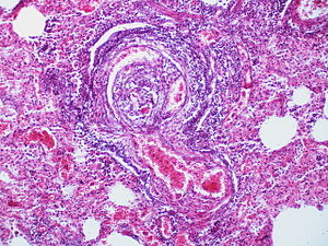
'''Pulmonary | [[Image:Angiomatoid (plexiform) and dilatation lesions (4348913976).jpg|thumb|right|300px|Plexiform lesion of the lung, a finding of pulmonary hypertension. (WC/Rosen)]] | ||
'''Pulmonary hypertension''' is bad stuff that arises from [[heart]] problems, an assortment of lung pathologies and some drugs. | |||
''Hypertension'', more generally, is dealt with in the ''[[hypertension]]'' article. | |||
==General classification== | ==General classification== | ||
| Line 7: | Line 10: | ||
===Non-secondary pulmonary hypertension=== | ===Non-secondary pulmonary hypertension=== | ||
Causes:<ref name=pmid16263465>{{cite journal |author=Bush A |title=Pulmonary hypertensive diseases |journal=Paediatr Respir Rev |volume=1 |issue=4 |pages=361-7 |year=2000 |month=December |pmid=16263465 |doi=10.1053/prrv.2000.0077 |url=}}</ref> | Causes:<ref name=pmid16263465>{{cite journal |author=Bush A |title=Pulmonary hypertensive diseases |journal=Paediatr Respir Rev |volume=1 |issue=4 |pages=361-7 |year=2000 |month=December |pmid=16263465 |doi=10.1053/prrv.2000.0077 |url=}}</ref> | ||
*Primary pulmonary hypertension. | *[[Primary pulmonary hypertension]]. | ||
*[[pulmonary embolism|Pulmonary embolic disease]] (thromboembolism, and non-thrombotic embolism). | *[[pulmonary embolism|Pulmonary embolic disease]] (thromboembolism, and non-thrombotic embolism). | ||
*Pulmonary capillary haemangiomatosis (PCH). | *[[Pulmonary capillary haemangiomatosis]] (PCH). | ||
*Pulmonary veno-occlusive disease (PVOD). | *[[Pulmonary veno-occlusive disease]] (PVOD). | ||
Notes: | Notes: | ||
| Line 25: | Line 28: | ||
===Microscopic=== | ===Microscopic=== | ||
*Like chronic pulmonary hypertension due to congenital heart disease but ''without'' the congenital heart disease.<ref name=dccpad/> | *Like chronic pulmonary hypertension due to [[congenital heart disease]] but ''without'' the congenital heart disease.<ref name=dccpad/> | ||
**Classified by ''Heath-Edwards classification'' (see below) into six grades. | **Classified by ''Heath-Edwards classification'' (see below) into six grades. | ||
==Pulmonary veno-occlusive disease | ====Images==== | ||
<gallery> | |||
* | Image:Angiomatoid_(plexiform)_and_dilatation_lesions_(4348914566).jpg | Plexiform lesion of the lung. (WC/Rosen) | ||
*Thrombosis - small veins & venules, particularily at the interlobular septae. | Image:Angiomatoid_(plexiform)_and_dilatation_lesions_(4348913976).jpg | Plexiform lesion of the lung. (WC/Rosen) | ||
Image:Angiomatoid (plexiform) and dilatation lesions (4348915198).jpg| Plexiform lesion of the lung. (WC/Rosen) | |||
</gallery> | |||
==Pulmonary veno-occlusive disease== | |||
*Abbreviated ''PVOD''. | |||
===General=== | |||
Clinical:<ref name=PPP393-6>{{Ref PPP|393-6}}</ref> | |||
*Gradual [[dyspnea]]. | |||
*+/-Non-productive cough. | |||
*+/-[[Clubbing]]. | |||
===Microscopic=== | |||
Features:<ref name=PPP393-6>{{Ref PPP|393-6}}</ref> | |||
*[[Thrombosis]] - small veins & venules, particularily at the interlobular septae. | |||
*Associated with mild homogenous peripheral interstitial fibrosis. | *Associated with mild homogenous peripheral interstitial fibrosis. | ||
| Line 70: | Line 88: | ||
**Grade 4: | **Grade 4: | ||
***Intima: '''"plexiform lesions"''' + fibrous & fibroelastic reaction, + cellular intimal reaction. | ***Intima: '''"plexiform lesions"''' + fibrous & fibroelastic reaction, + cellular intimal reaction. | ||
*****Plexiform lesions = multiple channels that are dilated, | *****Plexiform lesions = multiple channels that are dilated, associated with loss of elastic laminae; thought to arise at branch points due to aberrant WSS.<ref>[http://pathhsw5m54.ucsf.edu/overview/vessels.html http://pathhsw5m54.ucsf.edu/overview/vessels.html]</ref> | ||
***Media: generalized dilation +/- '''local "dilation lesions"'''. | ***Media: generalized dilation +/- '''local "dilation lesions"'''. | ||
***Micrographs: [http://pathhsw5m54.ucsf.edu/overview/vessels.html Plexiform lesions (ucsf.edu)], [http://www.pvrireview.org/viewimage.asp?img=PVRIReview_2009_1_1_34_44882_u6.jpg Plexiform lesions (pvrireview.org)]. | ***Micrographs: [http://pathhsw5m54.ucsf.edu/overview/vessels.html Plexiform lesions (ucsf.edu)], [http://www.pvrireview.org/viewimage.asp?img=PVRIReview_2009_1_1_34_44882_u6.jpg Plexiform lesions (pvrireview.org)]. | ||
Latest revision as of 18:13, 31 January 2022
Pulmonary hypertension is bad stuff that arises from heart problems, an assortment of lung pathologies and some drugs.
Hypertension, more generally, is dealt with in the hypertension article.
General classification
- Primary, i.e. primary pulmonary hypertension, or
- Secondary, e.g. due to congenital heart disease (like ventricular septal defect), interstitial pulmonary fibrosis.
Non-secondary pulmonary hypertension
Causes:[1]
- Primary pulmonary hypertension.
- Pulmonary embolic disease (thromboembolism, and non-thrombotic embolism).
- Pulmonary capillary haemangiomatosis (PCH).
- Pulmonary veno-occlusive disease (PVOD).
Notes:
- Some people consider PCH and PVOD to the be same thing.[2]
- Both have a poor prognosis.
- Clinically they present the same way.
- PVOD is based on case reports - it is extremely rare.[3]
Primary pulmonary hypertension
- AKA pulmonary plexogenic arteriopathy.[4]
General
- Familial PPH may be associated with BMPR2 mutations.[5]
Microscopic
- Like chronic pulmonary hypertension due to congenital heart disease but without the congenital heart disease.[4]
- Classified by Heath-Edwards classification (see below) into six grades.
Images
Pulmonary veno-occlusive disease
- Abbreviated PVOD.
General
Clinical:[6]
Microscopic
Features:[6]
- Thrombosis - small veins & venules, particularily at the interlobular septae.
- Associated with mild homogenous peripheral interstitial fibrosis.
DDx: chronic interstitial pneumonia.
Pulmonary capillary hemangiomatosis
- Abbreviated PCH.
General
- First reported in 1978 by Wagenvoort et al..[7]
Microscopic
Features:
- Proliferating and invasive capillaries.[8]
- Demonstrated by CD34 immunostaining.[2]
- Dilated capillaries[9][10] - key feature.
DDx:
- Passive congestion (PC).
- Differentiated by fact that PCH has multiple channels in alveolar wall (PC has only one).
Chronic pulmonary hypertension due to congenital heart disease
Heath-Edwards classification
Definition:[11]
- Six grades - based on intimal reaction and media of arteries and arterioles:
- Grade 1:
- Intima: no intimal reaction.
- Media: hypertrophied.
- Grade 2:
- Intima: cellular intimal reaction.
- Media: hypertrophied.
- Grade 3:
- Intima: fibrous & fibroelastic reaction + cellular intimal reaction.
- Media: hypertrophy +/- generalized dilation.
- Grade 4:
- Intima: "plexiform lesions" + fibrous & fibroelastic reaction, + cellular intimal reaction.
- Plexiform lesions = multiple channels that are dilated, associated with loss of elastic laminae; thought to arise at branch points due to aberrant WSS.[13]
- Media: generalized dilation +/- local "dilation lesions".
- Micrographs: Plexiform lesions (ucsf.edu), Plexiform lesions (pvrireview.org).
- Intima: "plexiform lesions" + fibrous & fibroelastic reaction, + cellular intimal reaction.
- Grade 5:
- Intima: as in Grade 4.
- Media: generalized dilation + local "dilation lesions" + pulmonary hemosiderosis.
- Grade 6:
- Intima: as in Grade 4.
- Media: generalized dilation + local "dilation lesions" + pulmonary hemosiderosis + necrotizing arteritis.
- Grade 1:
Notes:
- Bolded text - defining feature.
See also
References
- ↑ Bush A (December 2000). "Pulmonary hypertensive diseases". Paediatr Respir Rev 1 (4): 361-7. doi:10.1053/prrv.2000.0077. PMID 16263465.
- ↑ 2.0 2.1 Lantuéjoul S, Sheppard MN, Corrin B, Burke MM, Nicholson AG (July 2006). "Pulmonary veno-occlusive disease and pulmonary capillary hemangiomatosis: a clinicopathologic study of 35 cases". Am. J. Surg. Pathol. 30 (7): 850-7. doi:10.1097/01.pas.0000209834.69972.e5. PMID 16819327.
- ↑ Vevaina JR, Mark EJ (March 1988). "Thoracic hemangiomatosis masquerading as interstitial lung disease". Chest 93 (3): 657-9. PMID 3342678.
- ↑ 4.0 4.1 Lie JT, Silver MD. Diagnostic criteria of cardiovascular pathology: acquired diseases. ISBN 0-397-51630-4. PP.208-9.
- ↑ Online 'Mendelian Inheritance in Man' (OMIM) /600799
- ↑ 6.0 6.1 Leslie, Kevin O.; Wick, Mark R. (2004). Practical Pulmonary Pathology: A Diagnostic Approach (1st ed.). Churchill Livingstone. pp. 393-6. ISBN 978-0443066313.
- ↑ Wagenvoort CA, Beetstra A, Spijker J (November 1978). "Capillary haemangiomatosis of the lungs". Histopathology 2 (6): 401-6. PMID 730121.
- ↑ Tron V, Magee F, Wright JL, Colby T, Churg A (November 1986). "Pulmonary capillary hemangiomatosis". Hum. Pathol. 17 (11): 1144-50. PMID 3770733.
- ↑ MC August 2009.
- ↑ Leslie, Kevin O.; Wick, Mark R. (2004). Practical Pulmonary Pathology: A Diagnostic Approach (1st ed.). Churchill Livingstone. pp. 396-7. ISBN 978-0443066313.
- ↑ 11.0 11.1 HEATH D, EDWARDS JE (October 1958). "The pathology of hypertensive pulmonary vascular disease; a description of six grades of structural changes in the pulmonary arteries with special reference to congenital cardiac septal defects". Circulation 18 (4 Part 1): 533-47. PMID 13573570.
- ↑ Jaklitsch MT, Linden BC, Braunlin EA, Bolman RM, Foker JE (June 2001). "Open-lung biopsy guides therapy in children". Ann. Thorac. Surg. 71 (6): 1779-85. PMID 11426747.
- ↑ http://pathhsw5m54.ucsf.edu/overview/vessels.html


