Difference between revisions of "Epithelial-myoepithelial carcinoma"
Jump to navigation
Jump to search
(fix redirect) |
|||
| (6 intermediate revisions by the same user not shown) | |||
| Line 1: | Line 1: | ||
# | {{ Infobox diagnosis | ||
| Name = {{PAGENAME}} | |||
| Image = Epithelial-myoepithelial carcinoma - very high mag.jpg | |||
| Width = | |||
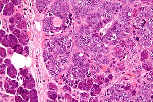
| Caption = Epithelial-myoepithelial carcinoma. [[H&E stain]]. | |||
| Micro = biphasic tumour with (1) epithelial layer, and (2) myoepithelial layer; variable architecture (solid, cystic, tubular, papillary); +/- spindle cells | |||
| Subtypes = | |||
| LMDDx = [[adenoid cystic carcinoma]] (tubular variant), [[pleomorphic adenoma]], tubular variant. | |||
| Stains = | |||
| IHC = CAM5.2 +ve (epithelial comp.), p63 +ve (myoepithelial comp.) | |||
| EM = | |||
| Molecular = | |||
| IF = | |||
| Gross = | |||
| Grossing = | |||
| Site = [[salivary gland]] - usually parotid gland | |||
| Assdx = | |||
| Syndromes = | |||
| Clinicalhx = | |||
| Signs = salivary gland mass | |||
| Symptoms = | |||
| Prevalence = very rare | |||
| Bloodwork = | |||
| Rads = | |||
| Endoscopy = | |||
| Prognosis = | |||
| Other = | |||
| ClinDDx = other [[salivary gland]] masses | |||
}} | |||
Epithelial-myoepithelial carcinoma, abbreviated '''EMCa''', is a rare malignant [[salivary gland]] tumour. | |||
Should '''not''' be confused with ''[[myoepithelial carcinoma]]''. | |||
==General== | |||
*Rare ~1% of salivary gland tumours.<ref name=pmid9568184>{{Cite journal | last1 = Tralongo | first1 = V. | last2 = Daniele | first2 = E. | title = Epithelial-myoepithelial carcinoma of the salivary glands: a review of literature. | journal = Anticancer Res | volume = 18 | issue = 1B | pages = 603-8 | month = | year = | doi = | PMID = 9568184 }}</ref> | |||
*Female:male = 1.5:1.<ref name=pmid17197918>{{Cite journal | last1 = Seethala | first1 = RR. | last2 = Barnes | first2 = EL. | last3 = Hunt | first3 = JL. | title = Epithelial-myoepithelial carcinoma: a review of the clinicopathologic spectrum and immunophenotypic characteristics in 61 tumors of the salivary glands and upper aerodigestive tract. | journal = Am J Surg Pathol | volume = 31 | issue = 1 | pages = 44-57 | month = Jan | year = 2007 | doi = 10.1097/01.pas.0000213314.74423.d8 | PMID = 17197918 }}</ref> | |||
*Usually older people - 50s or 60s. | |||
*Usually parotid gland ~ 60% of cases.<ref name=pmid17197918/> | |||
*Prognosis: usually good; 5-year and 10-year survival over 90% and 80% respectively.<ref name=pmid17197918/> | |||
Notes: | |||
*Most common malignant component in ''[[carcinoma ex pleomorphic adenoma]]''. | |||
*May be the same tumour as ''[[adenomyoepithelioma]] of the breast''.<ref name=pmid9769134>{{Cite journal | last1 = Seifert | first1 = G. | title = Are adenomyoepithelioma of the breast and epithelial-myoepithelial carcinoma of the salivary glands identical tumours? | journal = Virchows Arch | volume = 433 | issue = 3 | pages = 285-8 | month = Sep | year = 1998 | doi = | PMID = 9769134 }}</ref> | |||
*An analogous [[lung tumour]] exists - see ''[[epithelial-myoepithelial carcinoma of lung]].<ref>{{Cite journal | last1 = Nakashima | first1 = Y. | last2 = Morita | first2 = R. | last3 = Ui | first3 = A. | last4 = Iihara | first4 = K. | last5 = Yazawa | first5 = T. | title = Epithelial-myoepithelial carcinoma of the lung: a case report. | journal = Surg Case Rep | volume = 4 | issue = 1 | pages = 74 | month = Jul | year = 2018 | doi = 10.1186/s40792-018-0482-8 | PMID = 29987577 }}</ref> | |||
==Microscopic== | |||
Features: | |||
*Biphasic tumour:<ref name=pmid17197918/> | |||
*#Epithelial layer. | |||
*#Myoepithelial layer - '''key feature'''. | |||
*Architecture: variable (solid, cystic, tubular, papillary). | |||
*+/-Spindle cells. | |||
*Basement membrane-like material; may mimic adenoid cystic carcinoma. | |||
Notes: | |||
*Usually few mitoses. | |||
DDx: | |||
*[[Adenoid cystic carcinoma]] (tubular variant). | |||
*[[Pleomorphic adenoma]], tubular variant. | |||
**Has focal epithelial-myoepithelial carcinoma-like areas. | |||
===Images=== | |||
<gallery> | |||
Image:Epithelial-myoepithelial_carcinoma_-_intermed_mag.jpg | EMCa - intermed. mag. (WC/Nephron) | |||
Image:Epithelial-myoepithelial_carcinoma_-_high_mag.jpg | EMCa - high mag. (WC/Nephron) | |||
Image:Epithelial-myoepithelial carcinoma - very high mag.jpg | EMCa - very high mag. (WC/Nephron) | |||
</gallery> | |||
www: | |||
*[http://www.pathologyimagesinc.com/sgt-cytopath/epith-myoepith-ca/cytopathology/images-features/emc-rev-cyto1-18.jpg EMCa (pathologyimagesinc.com)].<ref>{{cite web |url=http://www.pathologyimagesinc.com/sgt-cytopath/epith-myoepith-ca/cytopathology/fs-emc-cytopath-feat.html |title=Cytopathologic Features of | |||
Epithelial-myoepithelial Carcinoma |last1= |first1= |last2= |first2= |date= |work= |publisher= |accessdate=January 18, 2011}}</ref> | |||
*[http://www.headandneckoncology.org/content/2/1/4 EMCa (headandneckoncology.org)]. | |||
==IHC== | |||
*CAM5.2 +ve -- epithelial component. | |||
*p63 +ve -- myoepithelial component. | |||
==See also== | |||
*[[Salivary glands]]. | |||
==References== | |||
{{Reflist|2}} | |||
[[Category:Diagnosis]] | |||
[[Category:Salivary gland]] | |||
[[Category:Head and neck pathology]] | |||
Latest revision as of 02:13, 19 March 2019
| Epithelial-myoepithelial carcinoma | |
|---|---|
| Diagnosis in short | |
 Epithelial-myoepithelial carcinoma. H&E stain. | |
|
| |
| LM | biphasic tumour with (1) epithelial layer, and (2) myoepithelial layer; variable architecture (solid, cystic, tubular, papillary); +/- spindle cells |
| LM DDx | adenoid cystic carcinoma (tubular variant), pleomorphic adenoma, tubular variant. |
| IHC | CAM5.2 +ve (epithelial comp.), p63 +ve (myoepithelial comp.) |
| Site | salivary gland - usually parotid gland |
|
| |
| Signs | salivary gland mass |
| Prevalence | very rare |
| Clin. DDx | other salivary gland masses |
Epithelial-myoepithelial carcinoma, abbreviated EMCa, is a rare malignant salivary gland tumour.
Should not be confused with myoepithelial carcinoma.
General
- Rare ~1% of salivary gland tumours.[1]
- Female:male = 1.5:1.[2]
- Usually older people - 50s or 60s.
- Usually parotid gland ~ 60% of cases.[2]
- Prognosis: usually good; 5-year and 10-year survival over 90% and 80% respectively.[2]
Notes:
- Most common malignant component in carcinoma ex pleomorphic adenoma.
- May be the same tumour as adenomyoepithelioma of the breast.[3]
- An analogous lung tumour exists - see epithelial-myoepithelial carcinoma of lung.[4]
Microscopic
Features:
- Biphasic tumour:[2]
- Epithelial layer.
- Myoepithelial layer - key feature.
- Architecture: variable (solid, cystic, tubular, papillary).
- +/-Spindle cells.
- Basement membrane-like material; may mimic adenoid cystic carcinoma.
Notes:
- Usually few mitoses.
DDx:
- Adenoid cystic carcinoma (tubular variant).
- Pleomorphic adenoma, tubular variant.
- Has focal epithelial-myoepithelial carcinoma-like areas.
Images
www:
IHC
- CAM5.2 +ve -- epithelial component.
- p63 +ve -- myoepithelial component.
See also
References
- ↑ Tralongo, V.; Daniele, E.. "Epithelial-myoepithelial carcinoma of the salivary glands: a review of literature.". Anticancer Res 18 (1B): 603-8. PMID 9568184.
- ↑ 2.0 2.1 2.2 2.3 Seethala, RR.; Barnes, EL.; Hunt, JL. (Jan 2007). "Epithelial-myoepithelial carcinoma: a review of the clinicopathologic spectrum and immunophenotypic characteristics in 61 tumors of the salivary glands and upper aerodigestive tract.". Am J Surg Pathol 31 (1): 44-57. doi:10.1097/01.pas.0000213314.74423.d8. PMID 17197918.
- ↑ Seifert, G. (Sep 1998). "Are adenomyoepithelioma of the breast and epithelial-myoepithelial carcinoma of the salivary glands identical tumours?". Virchows Arch 433 (3): 285-8. PMID 9769134.
- ↑ Nakashima, Y.; Morita, R.; Ui, A.; Iihara, K.; Yazawa, T. (Jul 2018). "Epithelial-myoepithelial carcinoma of the lung: a case report.". Surg Case Rep 4 (1): 74. doi:10.1186/s40792-018-0482-8. PMID 29987577.
- ↑ [http://www.pathologyimagesinc.com/sgt-cytopath/epith-myoepith-ca/cytopathology/fs-emc-cytopath-feat.html "Cytopathologic Features of Epithelial-myoepithelial Carcinoma"]. http://www.pathologyimagesinc.com/sgt-cytopath/epith-myoepith-ca/cytopathology/fs-emc-cytopath-feat.html. Retrieved January 18, 2011.


