Difference between revisions of "Diffuse astrocytoma, MYB- or MYBL-altered"
Jump to navigation
Jump to search
Jensflorian (talk | contribs) (Create template) |
Jensflorian (talk | contribs) (+ image) |
||
| (3 intermediate revisions by the same user not shown) | |||
| Line 1: | Line 1: | ||
{{ Infobox diagnosis | {{ Infobox diagnosis | ||
| Name = {{PAGENAME}} | | Name = {{PAGENAME}} | ||
| Image = | | Image = Commons 20220404 000.jpg | ||
| Width = | | Width = | ||
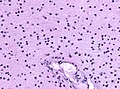
| Caption = Diffuse astrocytoma, MYB- or MYBL-altered [[H&E stain]]. | | Caption = Diffuse astrocytoma, MYB- or MYBL-altered [[H&E stain]]. | ||
| Line 30: | Line 30: | ||
| Tx = | | Tx = | ||
}} | }} | ||
'''Diffuse astrocytoma, MYB- or MYBL-altered''' is a low-grade pediatric [[astrocytoma]]. It is very rare. | '''Diffuse astrocytoma, MYB- or MYBL-altered''' is a low-grade, often pediatric-onset [[astrocytoma]]. It is very rare. | ||
==General== | ==General== | ||
* | * CNS WHO grade 1. | ||
* Entity introduced in 2021. <ref>{{cite journal |vauthors=Bale TA, Rosenblum MK |title=The 2021 WHO Classification of Tumors of the Central Nervous System: An update on pediatric low-grade gliomas and glioneuronal tumors |journal=Brain Pathol |volume= |issue= |pages=e13060 |date=February 2022 |pmid=35218102 |doi=10.1111/bpa.13060 |url=}}</ref> | |||
* Rare epilepsy-associated tumor. | |||
* Median time to surgery often exceeds more than 10 years. | |||
* No sex predilection. | |||
==Imaging== | ==Imaging== | ||
* T1 hypointense. | |||
* Non-enhancing. | |||
* Occasionally large cysts. | |||
==Gross== | ==Gross== | ||
* Soft, unencapsulated. | |||
* Grey to white. | |||
==Microscopic== | ==Microscopic== | ||
Features: | Features: | ||
* | * Diffusely growing. | ||
* Minimal incereased cell density. | |||
* Monomorphic, bland glial cells. | |||
* Entrapped neurons. | |||
* Usu. no mitotic activity | |||
* | |||
* | |||
* | |||
* | |||
Notes: | Notes: | ||
* | *Focally angiocentric pattern may be present. | ||
DDx | DDx | ||
| Line 64: | Line 65: | ||
===Images=== | ===Images=== | ||
<gallery> | <gallery> | ||
File:Commons 20220404 000.jpg|H&E | |||
</gallery> | </gallery> | ||
==Stains== | ==Stains== | ||
Features: | Features: | ||
*GFAP +ve | *GFAP +ve. | ||
*Olig2 -ve. | |||
*MAP2 -ve. | |||
*CD34 -ve. | |||
*ATRX retained. | |||
==Molecular== | ==Molecular== | ||
* | * Structural variant of MYB or MYBL1 (most common fusion partners: MAML2, MMP16 , PCDGHA1).<ref>{{cite journal |vauthors=Qaddoumi I, Orisme W, Wen J, Santiago T, Gupta K, Dalton JD, Tang B, Haupfear K, Punchihewa C, Easton J, Mulder H, Boggs K, Shao Y, Rusch M, Becksfort J, Gupta P, Wang S, Lee RP, Brat D, Peter Collins V, Dahiya S, George D, Konomos W, Kurian KM, McFadden K, Serafini LN, Nickols H, Perry A, Shurtleff S, Gajjar A, Boop FA, Klimo PD, Mardis ER, Wilson RK, Baker SJ, Zhang J, Wu G, Downing JR, Tatevossian RG, Ellison DW |title=Genetic alterations in uncommon low-grade neuroepithelial tumors: BRAF, FGFR1, and MYB mutations occur at high frequency and align with morphology |journal=Acta Neuropathol |volume=131 |issue=6 |pages=833–45 |date=June 2016 |pmid=26810070 |pmc=4866893 |doi=10.1007/s00401-016-1539-z |url=}}</ref> | ||
* IDH1/2 wildtype. | |||
* H3F3A wildtype. | |||
==Prognosis== | ==Prognosis== | ||
*Excellent ( | * Excellent (most cases are stable after surgery). | ||
==See also== | ==See also== | ||
Latest revision as of 07:50, 4 April 2022
Diffuse astrocytoma, MYB- or MYBL-altered is a low-grade, often pediatric-onset astrocytoma. It is very rare.
| Diffuse astrocytoma, MYB- or MYBL-altered | |
|---|---|
| Diagnosis in short | |
 Diffuse astrocytoma, MYB- or MYBL-altered H&E stain. | |
| LM DDx | Angiocentric glioma |
| IHC | GFAP +ve |
| Gross | soft, unencapsulated |
| Site | brain - usu. temporal |
|
| |
| Prevalence | common - esp. in children |
| Prognosis | good (WHO Grade I) |
General
- CNS WHO grade 1.
- Entity introduced in 2021. [1]
- Rare epilepsy-associated tumor.
- Median time to surgery often exceeds more than 10 years.
- No sex predilection.
Imaging
- T1 hypointense.
- Non-enhancing.
- Occasionally large cysts.
Gross
- Soft, unencapsulated.
- Grey to white.
Microscopic
Features:
- Diffusely growing.
- Minimal incereased cell density.
- Monomorphic, bland glial cells.
- Entrapped neurons.
- Usu. no mitotic activity
Notes:
- Focally angiocentric pattern may be present.
DDx
- Normal white matter
- Angiocentric glioma
Images
H&E
Stains
Features:
- GFAP +ve.
- Olig2 -ve.
- MAP2 -ve.
- CD34 -ve.
- ATRX retained.
Molecular
- Structural variant of MYB or MYBL1 (most common fusion partners: MAML2, MMP16 , PCDGHA1).[2]
- IDH1/2 wildtype.
- H3F3A wildtype.
Prognosis
- Excellent (most cases are stable after surgery).
See also
References
- ↑ "The 2021 WHO Classification of Tumors of the Central Nervous System: An update on pediatric low-grade gliomas and glioneuronal tumors". Brain Pathol: e13060. February 2022. doi:10.1111/bpa.13060. PMID 35218102.
- ↑ "Genetic alterations in uncommon low-grade neuroepithelial tumors: BRAF, FGFR1, and MYB mutations occur at high frequency and align with morphology". Acta Neuropathol 131 (6): 833–45. June 2016. doi:10.1007/s00401-016-1539-z. PMC 4866893. PMID 26810070. https://www.ncbi.nlm.nih.gov/pmc/articles/PMC4866893/.