Difference between revisions of "Nodal nevus"
Jump to navigation
Jump to search
(+infobox) |
(tweak) |
||
| (5 intermediate revisions by the same user not shown) | |||
| Line 1: | Line 1: | ||
{{ Infobox diagnosis | {{ Infobox diagnosis | ||
| Name = {{PAGENAME}} | | Name = {{PAGENAME}} | ||
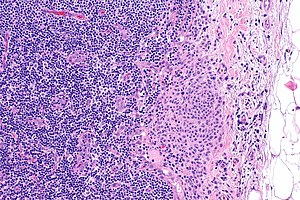
| Image = | | Image = Nodal nevus -- intermed mag.jpg | ||
| Width = | | Width = | ||
| Caption = | | Caption = | ||
| Synonyms = capsular nevus, benign nevus cells in lymph node | | Synonyms = capsular nevus, benign nevus cells in lymph node | ||
| Micro = | | Micro = bland nevus cells (lack prominent nucleoli, [[nuclear pleomorphism]], proliferative activity) | ||
| Subtypes = | | Subtypes = | ||
| LMDDx = [[malignant melanoma]] | | LMDDx = [[malignant melanoma]] | ||
| Line 29: | Line 29: | ||
| Other = | | Other = | ||
| ClinDDx = [[lymph node metastasis]] | | ClinDDx = [[lymph node metastasis]] | ||
| Tx = none | | Tx = none or follow-up | ||
}} | }} | ||
'''Nodal nevus''', also '''capsular nevus''', is benign [[lymph node]] | '''Nodal nevus''', also '''capsular nevus''', is benign [[lymph node]] finding that can be mistaken for [[malignant melanoma]].<ref name=pmid25081754>{{Cite journal | last1 = Lee | first1 = JJ. | last2 = Granter | first2 = SR. | last3 = Laga | first3 = AC. | last4 = Saavedra | first4 = AP. | last5 = Zhan | first5 = Q. | last6 = Guo | first6 = W. | last7 = Xu | first7 = S. | last8 = Murphy | first8 = GF. | last9 = Lian | first9 = CG. | title = 5-Hydroxymethylcytosine expression in metastatic melanoma versus nodal nevus in sentinel lymph node biopsies. | journal = Mod Pathol | volume = 28 | issue = 2 | pages = 218-29 | month = Feb | year = 2015 | doi = 10.1038/modpathol.2014.99 | PMID = 25081754 }}</ref> | ||
''Nevus in lymph node'' | '''Nevus in lymph node''' and '''benign nevus cells in lymph node''' redirect here. | ||
==General== | ==General== | ||
*[[Lymph node metastases]] from cutaneous melanoma are common and seen in approximately 20% of cases.<ref name=pmid28802493>{{Cite journal | last1 = Prieto | first1 = VG. | title = Sentinel Lymph Nodes in Cutaneous Melanoma. | journal = Clin Lab Med | volume = 37 | issue = 3 | pages = 417-430 | month = 09 | year = 2017 | doi = 10.1016/j.cll.2017.05.002 | PMID = 28802493 }}</ref> | *[[Lymph node metastases]] from cutaneous melanoma are common and seen in approximately 20% of cases.<ref name=pmid28802493>{{Cite journal | last1 = Prieto | first1 = VG. | title = Sentinel Lymph Nodes in Cutaneous Melanoma. | journal = Clin Lab Med | volume = 37 | issue = 3 | pages = 417-430 | month = 09 | year = 2017 | doi = 10.1016/j.cll.2017.05.002 | PMID = 28802493 }}</ref> | ||
* | *Nodal nevus may be confused with [[malignant melanoma]].<ref name=pmid27810606>{{Cite journal | last1 = Davis | first1 = J. | last2 = Patil | first2 = J. | last3 = Aydin | first3 = N. | last4 = Mishra | first4 = A. | last5 = Misra | first5 = S. | title = Capsular nevus versus metastatic malignant melanoma - a diagnostic dilemma. | journal = Int J Surg Case Rep | volume = 29 | issue = | pages = 20-24 | month = | year = 2016 | doi = 10.1016/j.ijscr.2016.10.040 | PMID = 27810606 }}</ref> | ||
==Gross== | ==Gross== | ||
| Line 53: | Line 53: | ||
DDx: | DDx: | ||
*[[Malignant melanoma]]. | *[[Malignant melanoma]]. | ||
===Images=== | |||
<gallery> | |||
Image: Nodal nevus -- very low mag.jpg | NN - very low mag. (WC) | |||
Image: Nodal nevus -- low mag.jpg | NN - low mag. (WC) | |||
Image: Nodal nevus -- intermed mag.jpg | NN - intermed. mag. (WC) | |||
Image: Nodal nevus -- high mag.jpg | NN - high mag. (WC) | |||
Image: Nodal nevus -- very high mag.jpg | NN - very high mag. (WC) | |||
</gallery> | |||
==IHC== | ==IHC== | ||
Latest revision as of 14:36, 16 October 2019
| Nodal nevus | |
|---|---|
| Diagnosis in short | |
 | |
|
| |
| Synonyms | capsular nevus, benign nevus cells in lymph node |
|
| |
| LM | bland nevus cells (lack prominent nucleoli, nuclear pleomorphism, proliferative activity) |
| LM DDx | malignant melanoma |
| IHC | S-100 +ve |
| Gross | usually not apparent |
| Site | lymph node |
|
| |
| Signs | none |
| Prevalence | rare |
| Prognosis | benign |
| Clin. DDx | lymph node metastasis |
| Treatment | none or follow-up |
Nodal nevus, also capsular nevus, is benign lymph node finding that can be mistaken for malignant melanoma.[1]
Nevus in lymph node and benign nevus cells in lymph node redirect here.
General
- Lymph node metastases from cutaneous melanoma are common and seen in approximately 20% of cases.[2]
- Nodal nevus may be confused with malignant melanoma.[3]
Gross
- Usually not apparent.
Microscopic
Features:
- Nevus cells with bland cytomorphology.
- Lack size variation, nucleoli.
- Non-proliferative.
Note:
- No standardized criteria.[3]
DDx:
Images
IHC
Features:[3]
- S-100 +ve.
References
- ↑ Lee, JJ.; Granter, SR.; Laga, AC.; Saavedra, AP.; Zhan, Q.; Guo, W.; Xu, S.; Murphy, GF. et al. (Feb 2015). "5-Hydroxymethylcytosine expression in metastatic melanoma versus nodal nevus in sentinel lymph node biopsies.". Mod Pathol 28 (2): 218-29. doi:10.1038/modpathol.2014.99. PMID 25081754.
- ↑ Prieto, VG. (09 2017). "Sentinel Lymph Nodes in Cutaneous Melanoma.". Clin Lab Med 37 (3): 417-430. doi:10.1016/j.cll.2017.05.002. PMID 28802493.
- ↑ 3.0 3.1 3.2 Davis, J.; Patil, J.; Aydin, N.; Mishra, A.; Misra, S. (2016). "Capsular nevus versus metastatic malignant melanoma - a diagnostic dilemma.". Int J Surg Case Rep 29: 20-24. doi:10.1016/j.ijscr.2016.10.040. PMID 27810606.


