Difference between revisions of "Polarization"
Jump to navigation
Jump to search


| (2 intermediate revisions by the same user not shown) | |||
| Line 2: | Line 2: | ||
[[Image:Acquired_cystic_disease-associated_renal_cell_carcinoma_-2a--_intermed_mag.gif|thumb|right|[[Acquired cystic disease-associated renal cell carcinoma]] with non-polarized light and polarized light to highlight the oxylate crystals. [[H&E stain]]. (WC/Nephron)]] | [[Image:Acquired_cystic_disease-associated_renal_cell_carcinoma_-2a--_intermed_mag.gif|thumb|right|[[Acquired cystic disease-associated renal cell carcinoma]] with non-polarized light and polarized light to highlight the oxylate crystals. [[H&E stain]]. (WC/Nephron)]] | ||
'''Polarization''', formally '''light polarization''', in [[pathology]] refers to a technique used in [[light microscopy]] that makes use of polarized light. | '''Polarization''', formally '''light polarization''', in [[pathology]] refers to a technique used in [[light microscopy]] that makes use of polarized light. | ||
==General== | |||
*Polarization is a useful characteristic as it is only seen in handful of pathologies. | |||
*In some cases the colour and histomorphology can be specific enough to be diagnostic. | |||
==Notable things that polarize== | ==Notable things that polarize== | ||
*[[Amyloid]] - apple-green birefringence.<ref name=pmid26485243>{{Cite journal | last1 = Cornejo | first1 = KM. | last2 = Lagana | first2 = FJ. | last3 = Deng | first3 = A. | title = Nodular amyloidosis derived from keratinocytes: an unusual type of primary localized cutaneous nodular amyloidosis. | journal = Am J Dermatopathol | volume = 37 | issue = 11 | pages = e129-33 | month = Nov | year = 2015 | doi = 10.1097/DAD.0000000000000307 | PMID = 26485243 }}</ref> | *[[Amyloid]] - apple-green birefringence.<ref name=pmid26485243>{{Cite journal | last1 = Cornejo | first1 = KM. | last2 = Lagana | first2 = FJ. | last3 = Deng | first3 = A. | title = Nodular amyloidosis derived from keratinocytes: an unusual type of primary localized cutaneous nodular amyloidosis. | journal = Am J Dermatopathol | volume = 37 | issue = 11 | pages = e129-33 | month = Nov | year = 2015 | doi = 10.1097/DAD.0000000000000307 | PMID = 26485243 }}</ref> | ||
*Oxylate crystals | *Oxylate crystals. | ||
**[[Ethylene glycol poisoning]].<ref name=pmid19874660>{{Cite journal | last1 = Rosano | first1 = TG. | last2 = Swift | first2 = TA. | last3 = Kranick | first3 = CJ. | last4 = Sikirica | first4 = M. | title = Ethylene glycol and glycolic acid in postmortem blood from fatal poisonings. | journal = J Anal Toxicol | volume = 33 | issue = 8 | pages = 508-13 | month = Oct | year = 2009 | doi = | PMID = 19874660 }}</ref> | |||
**[[Acquired cystic disease-associated renal cell carcinoma]]. | |||
**Benign [[breast calcifications]].<ref name=pmid10097726>{{Cite journal | last1 = Ozer | first1 = E. | last2 = Canda | first2 = T. | last3 = Balci | first3 = P. | last4 = Gökçe | first4 = O. | title = Calcium oxalate crystals in benign cyst fluid from the breast. A case report. | journal = Acta Cytol | volume = 43 | issue = 2 | pages = 281-4 | month = | year = | doi = | PMID = 10097726 }}</ref> | |||
*[[Gout]] crystals - negatively birefringent, yellow when aligned, needle-shaped.<ref name=Ref_TN2005>{{Ref TN2005| RH6}}</ref> | *[[Gout]] crystals - negatively birefringent, yellow when aligned, needle-shaped.<ref name=Ref_TN2005>{{Ref TN2005| RH6}}</ref> | ||
*[[Pseudogout]] crystals - positively birefringent, blue when aligned, rhomboid-shaped.<ref name=Ref_TN2005>{{Ref TN2005| RH6}}</ref> | *[[Pseudogout]] crystals - positively birefringent, blue when aligned, rhomboid-shaped.<ref name=Ref_TN2005>{{Ref TN2005| RH6}}</ref> | ||
| Line 18: | Line 25: | ||
==References== | ==References== | ||
{{Reflist| | {{Reflist|2}} | ||
==External links== | |||
*[https://en.wikipedia.org/wiki/Birefringence Birefringence (wikipedia.org)]. | |||
[[Category:Basics]] | [[Category:Basics]] | ||
Latest revision as of 01:22, 25 April 2016

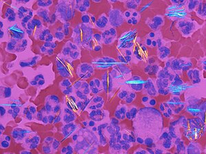
Crystals (gout) and blood cells in polarized light. (WC/Gabriel Caponetti)

Acquired cystic disease-associated renal cell carcinoma with non-polarized light and polarized light to highlight the oxylate crystals. H&E stain. (WC/Nephron)
Polarization, formally light polarization, in pathology refers to a technique used in light microscopy that makes use of polarized light.
General
- Polarization is a useful characteristic as it is only seen in handful of pathologies.
- In some cases the colour and histomorphology can be specific enough to be diagnostic.
Notable things that polarize
- Amyloid - apple-green birefringence.[1]
- Oxylate crystals.
- Gout crystals - negatively birefringent, yellow when aligned, needle-shaped.[4]
- Pseudogout crystals - positively birefringent, blue when aligned, rhomboid-shaped.[4]
Some other things that polarize
- Dirt.
- Collagen.
See also
References
- ↑ Cornejo, KM.; Lagana, FJ.; Deng, A. (Nov 2015). "Nodular amyloidosis derived from keratinocytes: an unusual type of primary localized cutaneous nodular amyloidosis.". Am J Dermatopathol 37 (11): e129-33. doi:10.1097/DAD.0000000000000307. PMID 26485243.
- ↑ Rosano, TG.; Swift, TA.; Kranick, CJ.; Sikirica, M. (Oct 2009). "Ethylene glycol and glycolic acid in postmortem blood from fatal poisonings.". J Anal Toxicol 33 (8): 508-13. PMID 19874660.
- ↑ Ozer, E.; Canda, T.; Balci, P.; Gökçe, O.. "Calcium oxalate crystals in benign cyst fluid from the breast. A case report.". Acta Cytol 43 (2): 281-4. PMID 10097726.
- ↑ 4.0 4.1 Yeung, J.C.; Leonard, Blair J. N. (2005). The Toronto Notes 2005 - Review for the MCCQE and Comprehensive Medical Reference (2005 ed.). The Toronto Notes Inc. for Medical Students Inc.. pp. RH6. ISBN 978-0968592854.