Difference between revisions of "Serous borderline tumour"
Jump to navigation
Jump to search
m |
|||
| (7 intermediate revisions by the same user not shown) | |||
| Line 1: | Line 1: | ||
{{ Infobox diagnosis | |||
| Name = {{PAGENAME}} | |||
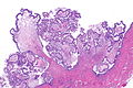
| Image = Serous borderline tumour with micropapillary pattern -- intermed mag.jpg | |||
| Width = | |||
| Caption = Serous borderline tumour with a micropapillary pattern. [[H&E stain]]. | |||
| Synonyms = | |||
| Micro = cuboidal to columnar epithelium with mild to moderate atypia, no invasion (see below), "sparse" mitoses ,+/-[[psammoma bodies]], +/-micropapillary architecture (often described as a ''medusa head'' pattern) | |||
| Subtypes = serous borderline tumour (SBT) with micropapillary pattern, typical SBT ''or'' SBT not otherwise specified | |||
| LMDDx = [[serous cystadenoma of the ovary|serous cystadenoma]], [[serous carcinoma of the ovary]], [[clear cell carcinoma of the ovary]] | |||
| Stains = | |||
| IHC = | |||
| EM = | |||
| Molecular = | |||
| IF = | |||
| Gross = | |||
| Grossing = | |||
| Staging = | |||
| Site = [[ovary]], [[uterine tube]] - see ''[[ovarian tumours]]'' | |||
| Assdx = | |||
| Syndromes = | |||
| Clinicalhx = | |||
| Signs = | |||
| Symptoms = | |||
| Prevalence = uncommon | |||
| Bloodwork = | |||
| Rads = | |||
| Endoscopy = | |||
| Prognosis = usually benign, need follow-up | |||
| Other = | |||
| ClinDDx = other [[ovarian tumours]] (benign and malignant) | |||
| Tx = excision, follow-up | |||
}} | |||
'''Serous borderline tumour''' is a Muellerian epithelial [[ovarian tumour]] with a behaviour that borders on [[malignant]]. | '''Serous borderline tumour''' is a Muellerian epithelial [[ovarian tumour]] with a behaviour that borders on [[malignant]]. | ||
| Line 12: | Line 44: | ||
Features:<ref name=Ref_GP399>{{Ref GP|399}}</ref> | Features:<ref name=Ref_GP399>{{Ref GP|399}}</ref> | ||
*Cuboidal to columnar epithelium with mild to moderate atypia. | *Cuboidal to columnar epithelium with mild to moderate atypia. | ||
* | *Non-invasive. | ||
*"Sparse" mitoses. | *"Sparse" mitoses. | ||
*+/-[[Psammoma bodies]]. | *+/-[[Psammoma bodies]]. | ||
| Line 21: | Line 53: | ||
**Invasive cells are "pink", i.e. have abundant eosinophilic cytoplasm,<ref name=Ref_GP399/>; also, cells usu. large (~2-3x size of non-invasive component), and typically have an enlarged nucleus (~2x non-invasive component). | **Invasive cells are "pink", i.e. have abundant eosinophilic cytoplasm,<ref name=Ref_GP399/>; also, cells usu. large (~2-3x size of non-invasive component), and typically have an enlarged nucleus (~2x non-invasive component). | ||
*[[Clear cell carcinoma of the ovary]] - classically associated with [[endometriosis]], have simpler, smaller papillae without branching. | *[[Clear cell carcinoma of the ovary]] - classically associated with [[endometriosis]], have simpler, smaller papillae without branching. | ||
*[[Serous cystadenoma of the ovary]]. | |||
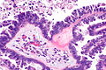
Images: | ===Images=== | ||
<gallery> | |||
Image: Serous borderline tumour with micropapillary pattern -- intermed mag.jpg -- very very low mag.jpg | SBTMP - very very low mag. | |||
Image: Serous borderline tumour with micropapillary pattern -- very low mag.jpg | SBTMP - very low mag. | |||
Image: Serous borderline tumour with micropapillary pattern -- low mag.jpg | SBTMP - low mag. | |||
Image: Serous borderline tumour with micropapillary pattern -- intermed mag.jpg | SBTMP - intermed. mag. | |||
Image: Serous borderline tumour with micropapillary pattern -- high mag.jpg | SBTMP - high mag. | |||
Image: Serous borderline tumour with micropapillary pattern - a -- very low mag.jpg | SBTMP - very low mag. | |||
Image: Serous borderline tumour with micropapillary pattern - a -- low mag.jpg | SBTMP - low mag. | |||
Image: Serous borderline tumour with micropapillary pattern - a -- intermed mag.jpg | SBTMP - intermed. mag. | |||
Image: Serous borderline tumour with micropapillary pattern - a -- high mag.jpg | SBTMP - high mag. | |||
Image: Serous borderline tumour with micropapillary pattern - a2 -- high mag.jpg | SBTMP - high mag. | |||
Image: Serous borderline tumour with micropapillary pattern - a3 -- high mag.jpg | SBTMP - high mag. | |||
Image: Serous borderline tumour with micropapillary pattern - a -- very high mag.jpg | SBTMP - very high mag. | |||
</gallery> | |||
====www==== | |||
*[http://pubs.rsna.org/na101/home/literatum/publisher/rsna/journals/content/radiographics/2005/radiographics.2005.25.issue-6/rg.256055015/production/images/medium/g05nv16c4x.jpeg Serous ovarian LMP tumour (radiographics.rsna.org)].<ref name=pmid16284143>{{Cite journal | last1 = Burkholz | first1 = KJ. | last2 = Wood | first2 = BP. | last3 = Zuppan | first3 = C. | title = Best cases from the AFIP: Borderline papillary serous tumor of the right ovary. | journal = Radiographics | volume = 25 | issue = 6 | pages = 1689-92 | month = | year = | doi = 10.1148/rg.256055015 | PMID = 16284143 }}</ref> | *[http://pubs.rsna.org/na101/home/literatum/publisher/rsna/journals/content/radiographics/2005/radiographics.2005.25.issue-6/rg.256055015/production/images/medium/g05nv16c4x.jpeg Serous ovarian LMP tumour (radiographics.rsna.org)].<ref name=pmid16284143>{{Cite journal | last1 = Burkholz | first1 = KJ. | last2 = Wood | first2 = BP. | last3 = Zuppan | first3 = C. | title = Best cases from the AFIP: Borderline papillary serous tumor of the right ovary. | journal = Radiographics | volume = 25 | issue = 6 | pages = 1689-92 | month = | year = | doi = 10.1148/rg.256055015 | PMID = 16284143 }}</ref> | ||
| Line 33: | Line 82: | ||
<pre> | <pre> | ||
Cyst and Right Fallopian Tube, Excision: | Cyst and Right Fallopian Tube, Excision: | ||
- SEROUS BORDERLINE TUMOUR with micropapillary architecture, see comment. | |||
- Fallopian tube within normal limits. | |||
- NEGATIVE for evidence of invasion. | |||
Comment: | Comment: | ||
Latest revision as of 11:25, 29 March 2016
| Serous borderline tumour | |
|---|---|
| Diagnosis in short | |
 Serous borderline tumour with a micropapillary pattern. H&E stain. | |
|
| |
| LM | cuboidal to columnar epithelium with mild to moderate atypia, no invasion (see below), "sparse" mitoses ,+/-psammoma bodies, +/-micropapillary architecture (often described as a medusa head pattern) |
| Subtypes | serous borderline tumour (SBT) with micropapillary pattern, typical SBT or SBT not otherwise specified |
| LM DDx | serous cystadenoma, serous carcinoma of the ovary, clear cell carcinoma of the ovary |
| Site | ovary, uterine tube - see ovarian tumours |
|
| |
| Prevalence | uncommon |
| Prognosis | usually benign, need follow-up |
| Clin. DDx | other ovarian tumours (benign and malignant) |
| Treatment | excision, follow-up |
Serous borderline tumour is a Muellerian epithelial ovarian tumour with a behaviour that borders on malignant.
It is also known as serous tumour of low malignant potential, abbreviated SLMP.[1][2]
Serous ovarian tumour of low malignant potential redirects here.[2]
General
- Usually benign.
- Require long term follow-up.
Microscopic
Features:[3]
- Cuboidal to columnar epithelium with mild to moderate atypia.
- Non-invasive.
- "Sparse" mitoses.
- +/-Psammoma bodies.
- +/-Micropapillary architecture - often described as a medusa head pattern.
DDx:
- Serous carcinoma of the ovary - focus a with stromal invasion >5mm (linear measurement) or > 10 mm2 (area).[3]
- Invasive cells are "pink", i.e. have abundant eosinophilic cytoplasm,[3]; also, cells usu. large (~2-3x size of non-invasive component), and typically have an enlarged nucleus (~2x non-invasive component).
- Clear cell carcinoma of the ovary - classically associated with endometriosis, have simpler, smaller papillae without branching.
- Serous cystadenoma of the ovary.
Images
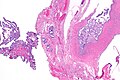
SBTMP - very very low mag.
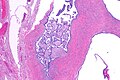
SBTMP - very low mag.
SBTMP - low mag.
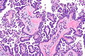
SBTMP - intermed. mag.

SBTMP - high mag.
SBTMP - very low mag.

SBTMP - low mag.

SBTMP - intermed. mag.

SBTMP - high mag.
SBTMP - high mag.
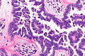
SBTMP - high mag.
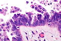
SBTMP - very high mag.


www
Subclassification
Typical subdivided into:[5]
- Micropapillary serous borderline tumour.
- Typical serous borderline tumour (SBOT).
Sign out
Cyst and Right Fallopian Tube, Excision:
- SEROUS BORDERLINE TUMOUR with micropapillary architecture, see comment.
- Fallopian tube within normal limits.
- NEGATIVE for evidence of invasion.

Comment:
The lesion appears to be confined to a cystic structure.
See also
References
- ↑ Seidman, JD.; Kurman, RJ. (May 2000). "Ovarian serous borderline tumors: a critical review of the literature with emphasis on prognostic indicators.". Hum Pathol 31 (5): 539-57. PMID 10836293.
- ↑ 2.0 2.1 Dietel, M.; Hauptmann, S. (May 2000). "Serous tumors of low malignant potential of the ovary. 1. Diagnostic pathology.". Virchows Arch 436 (5): 403-12. PMID 10881733.
- ↑ 3.0 3.1 3.2 Nucci, Marisa R.; Oliva, Esther (2009). Gynecologic Pathology: A Volume in Foundations in Diagnostic Pathology Series (1st ed.). Churchill Livingstone. pp. 399. ISBN 978-0443069208.
- ↑ Burkholz, KJ.; Wood, BP.; Zuppan, C.. "Best cases from the AFIP: Borderline papillary serous tumor of the right ovary.". Radiographics 25 (6): 1689-92. doi:10.1148/rg.256055015. PMID 16284143.
- ↑ Park, JY.; Kim, DY.; Kim, JH.; Kim, YM.; Kim, KR.; Kim, YT.; Nam, JH. (Dec 2011). "Micropapillary pattern in serous borderline ovarian tumors: does it matter?". Gynecol Oncol 123 (3): 511-6. doi:10.1016/j.ygyno.2011.08.008. PMID 21917305.