Difference between revisions of "Apocrine metaplasia of the breast"
Jump to navigation
Jump to search
| (2 intermediate revisions by the same user not shown) | |||
| Line 9: | Line 9: | ||
*Increased number of mitochondria. | *Increased number of mitochondria. | ||
**In other body sites this has different names, e.g. ''Hurthle cell change'' (thyroid), ''[[oncocytoma|oncocytic]] change'' (kidney). | **In other body sites this has different names, e.g. ''Hurthle cell change'' (thyroid), ''[[oncocytoma|oncocytic]] change'' (kidney). | ||
==Gross== | |||
*Blue dome cysts.<ref name=pmid25610180>{{Cite journal | last1 = Rodrigues | first1 = G. | last2 = Prabhu | first2 = R. | last3 = Nair | first3 = R. | last4 = Thinda | first4 = R. | title = A blue-domed cyst of bloodgood. | journal = Eurasian J Med | volume = 43 | issue = 2 | pages = 132 | month = Aug | year = 2011 | doi = 10.5152/eajm.2011.30 | PMID = 25610180 }}</ref> | |||
==Microscopic== | ==Microscopic== | ||
| Line 19: | Line 22: | ||
**Prominent nuclear membrane. | **Prominent nuclear membrane. | ||
**Prominent, often single nucleolus. | **Prominent, often single nucleolus. | ||
*+/-Luminal papillary tufts.<ref>URL: [http://www.breastpathology.info/Benign%20proliferative%20disease.html http://www.breastpathology.info/Benign%20proliferative%20disease.html]. Accessed on: May 9, 2016.</ref> | |||
Note: | Note: | ||
| Line 24: | Line 28: | ||
===Images=== | ===Images=== | ||
====Case==== | |||
<gallery> | |||
Image: Apocrine metaplasia -- low mag.jpg | AM - low mag. | |||
Image: Apocrine metaplasia -- intermed mag.jpg | AM - intermed. mag. | |||
Image: Apocrine metaplasia -- high mag.jpg | AM - high mag. | |||
Image: Apocrine metaplasia -- very high mag.jpg | AM - very high mag. | |||
Image: Apocrine metaplasia - alt -- very high mag.jpg | AM - very high mag. | |||
</gallery> | |||
====Others==== | |||
<gallery> | <gallery> | ||
Image:Fibrocystic_change_-_very_high_mag.jpg | FCC with apocrine metaplasia (right bottom of image) - high mag. (WC/Nephron). | Image:Fibrocystic_change_-_very_high_mag.jpg | FCC with apocrine metaplasia (right bottom of image) - high mag. (WC/Nephron). | ||
Latest revision as of 21:36, 9 May 2016
Apocrine metaplasia of the breast, also apocrine metaplasia, is a benign change in the breast without increased risk of malignancy.
General
- Benign/not significant. Can be considered to be pretty wallpaper in the house of breast pathology.
- Very common in adults.
- Apocrine lesions as a group are usually benign, some pre-neoplastic and some malignant.[1]
Etiology
- Increased number of mitochondria.
- In other body sites this has different names, e.g. Hurthle cell change (thyroid), oncocytic change (kidney).
Gross
- Blue dome cysts.[2]
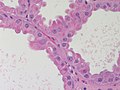
Microscopic
Features:
- Eosinophilic cytoplasm - key feature.
- Voluminous pink cytoplasm.
- Apocrine snouts may be present.
- Small protrusiona at the apical aspect of the cell (composed of cytoplasm and plasma membrane).
- Central round nucleus
- Prominent nuclear membrane.
- Prominent, often single nucleolus.
- +/-Luminal papillary tufts.[3]
Note:
- Apocrine changes, i.e. cytoplasmic eosinophilia, can appear in malignant tumours; eosinophilia doesn't make something benign.
Images
Case
- Apocrine metaplasia -- low mag.jpg
AM - low mag.
- Apocrine metaplasia -- intermed mag.jpg
AM - intermed. mag.
- Apocrine metaplasia -- high mag.jpg
AM - high mag.
- Apocrine metaplasia -- very high mag.jpg
AM - very high mag.
- Apocrine metaplasia - alt -- very high mag.jpg
AM - very high mag.
Others
- Fibrocystic change - very high mag.jpg
FCC with apocrine metaplasia (right bottom of image) - high mag. (WC/Nephron).
Breast - Apocrine Change - high power (SKB)

Sign out
- Typically not reported.
See also
References
- ↑ Wells, CA.; El-Ayat, GA. (Dec 2007). "Non-operative breast pathology: apocrine lesions.". J Clin Pathol 60 (12): 1313-20. doi:10.1136/jcp.2006.040626. PMID 18042688.
- ↑ Rodrigues, G.; Prabhu, R.; Nair, R.; Thinda, R. (Aug 2011). "A blue-domed cyst of bloodgood.". Eurasian J Med 43 (2): 132. doi:10.5152/eajm.2011.30. PMID 25610180.
- ↑ URL: http://www.breastpathology.info/Benign%20proliferative%20disease.html. Accessed on: May 9, 2016.