Difference between revisions of "Renal hybrid oncocytic/chromophobe tumour"
Jump to navigation
Jump to search
| (13 intermediate revisions by the same user not shown) | |||
| Line 1: | Line 1: | ||
{{ Infobox diagnosis | {{ Infobox diagnosis | ||
| Name = {{PAGENAME}} | | Name = {{PAGENAME}} | ||
| Image = | | Image = Hybrid tumour of the kidney -- high mag.jpg | ||
| Width = | | Width = | ||
| Caption = | | Caption = Hybrid oncocytic/chromophobe tumour of the kidney. [[H&E stain]]. | ||
| Synonyms = | | Synonyms = hybrid tumour | ||
| Micro = features of [[renal oncocytoma]] and [[chromophobe renal cell carcinoma]] | | Micro = features of [[renal oncocytoma]] and [[chromophobe renal cell carcinoma]] - varies by subtype | ||
| Subtypes = | | Subtypes = as per Hes ''et al.'': (1) [[collision tumour]]-type, (2) renal oncocytoma with scattered chromophobe cells-type, (3) large eosinophilic cells with intracytoplasmic vacuoles-type | ||
| LMDDx = [[renal oncocytoma]], [[chromophobe renal cell carcinoma]], [[renal cell carcinoma, unclassified]], other [[renal tumours with eosinophilic cytoplasm]] | | LMDDx = [[renal oncocytoma]], [[chromophobe renal cell carcinoma]], [[renal cell carcinoma, unclassified]], [[SDH-deficient renal cell carcinoma]], other [[renal tumours with eosinophilic cytoplasm]] | ||
| Stains = Hale's colloidal iron +ve | | Stains = Hale's colloidal iron +ve | ||
| IHC = CD117 +ve, CK7 +ve (variable) | | IHC = CD117 +ve, CK7 +ve (variable) | ||
| Line 14: | Line 14: | ||
| IF = | | IF = | ||
| Gross = | | Gross = | ||
| Grossing = | | Grossing = [[partial nephrectomy grossing]], [[total nephrectomy for tumour grossing]] | ||
| Site = [[kidney]] - see ''[[kidney tumours]]'' | | Site = [[kidney]] - see ''[[kidney tumours]]'' | ||
| Assdx = | | Assdx = | ||
| Line 40: | Line 40: | ||
*Sporadic. | *Sporadic. | ||
*[[Birt–Hogg–Dubé syndrome]]. | *[[Birt–Hogg–Dubé syndrome]]. | ||
*Renal oncocytosis. | *[[Renal oncocytosis]]. | ||
==Microscopic== | ==Microscopic== | ||
| Line 47: | Line 47: | ||
#* Different fields viewed in isolation would be compatible with the different diagnoses. Tumour component do not intermingle. | #* Different fields viewed in isolation would be compatible with the different diagnoses. Tumour component do not intermingle. | ||
# Renal oncocytoma with scattered chromophobe cells. | # Renal oncocytoma with scattered chromophobe cells. | ||
# Large eosinophilic cell with intracytoplasmic vacuoles. | # Large eosinophilic cell with intracytoplasmic vacuoles - this is the evolving entity ''[[eosinophilic vacuolated tumour]]''. | ||
#* Prominent nucleoli ([[ISUP nucleolar grade]] 3). | #* Prominent nucleoli ([[ISUP nucleolar grade]] 3). | ||
#* Perinuclear halos (occasional). | #* Perinuclear halos (occasional). | ||
| Line 55: | Line 55: | ||
*[[Renal oncocytoma]] - may have limited chromophobe-like areas (<=5% of tumour).<ref name=pmid21166703>{{Cite journal | last1 = Trpkov | first1 = K. | last2 = Yilmaz | first2 = A. | last3 = Uzer | first3 = D. | last4 = Dishongh | first4 = KM. | last5 = Quick | first5 = CM. | last6 = Bismar | first6 = TA. | last7 = Gokden | first7 = N. | title = Renal oncocytoma revisited: a clinicopathological study of 109 cases with emphasis on problematic diagnostic features. | journal = Histopathology | volume = 57 | issue = 6 | pages = 893-906 | month = Dec | year = 2010 | doi = 10.1111/j.1365-2559.2010.03726.x | PMID = 21166703 }}</ref> | *[[Renal oncocytoma]] - may have limited chromophobe-like areas (<=5% of tumour).<ref name=pmid21166703>{{Cite journal | last1 = Trpkov | first1 = K. | last2 = Yilmaz | first2 = A. | last3 = Uzer | first3 = D. | last4 = Dishongh | first4 = KM. | last5 = Quick | first5 = CM. | last6 = Bismar | first6 = TA. | last7 = Gokden | first7 = N. | title = Renal oncocytoma revisited: a clinicopathological study of 109 cases with emphasis on problematic diagnostic features. | journal = Histopathology | volume = 57 | issue = 6 | pages = 893-906 | month = Dec | year = 2010 | doi = 10.1111/j.1365-2559.2010.03726.x | PMID = 21166703 }}</ref> | ||
*[[Chromophobe renal cell carcinoma]], eosinophilic variant. | *[[Chromophobe renal cell carcinoma]], eosinophilic variant. | ||
*[[SDH-deficient renal cell carcinoma]] - lower nuclear grade, not nested. | |||
*[[Eosinophilic vacuolated tumour]] - evolving entity. | |||
*Other [[renal tumours with eosinophilic cytoplasm]]. | *Other [[renal tumours with eosinophilic cytoplasm]]. | ||
*[[Renal cell carcinoma, unclassified]]. | *[[Renal cell carcinoma, unclassified]]. | ||
===Images=== | |||
====Case 1==== | |||
<gallery> | |||
Image: Hybrid tumour of the kidney -- low mag.jpg | HOCT - low mag. (WC) | |||
Image: Hybrid tumour of the kidney -- intermed mag.jpg | HOCT - intermed. mag. (WC) | |||
Image: Hybrid tumour of the kidney -- high mag.jpg | HOCT - high mag. (WC) | |||
</gallery> | |||
====Case 2==== | |||
<gallery> | |||
Image: Renal hybrid tumour - 2 -- intermed mag.jpg | HOCT - intermed. mag. (WC) | |||
Image: Renal hybrid tumour - 2 -- high mag.jpg | HOCT - high mag. (WC) | |||
Image: Renal hybrid tumour - 2 -- very high mag.jpg | HOCT - very high mag. (WC) | |||
Image: Renal hybrid tumour - nests - 2 -- intermed mag.jpg | HOCT - intermed. mag. (WC) | |||
Image: Renal hybrid tumour - nests - 2 -- high mag.jpg | HOCT - high mag. (WC) | |||
Image: Renal hybrid tumour - nests - 2 -- very high mag.jpg | HOCT - very high mag. (WC) | |||
</gallery> | |||
==Stains== | ==Stains== | ||
| Line 75: | Line 95: | ||
==Molecular== | ==Molecular== | ||
*No features characteristic of [[chromophobe RCC]] on array-CGH analysis.<ref name=pmid23708994/> | *No features characteristic of [[chromophobe RCC]] on array-CGH analysis.<ref name=pmid23708994/> | ||
==Sign out== | |||
<pre> | |||
Left Kidney, Partial Nephrectomy: | |||
- Renal tumour with eosinophilic cytoplasm of undetermined malignant potential | |||
in keeping with the so called "hybrid oncocytic/chromophobe tumour", see comment. | |||
-- Resection margins clear. | |||
-- Tumour limited to kidney. | |||
Comment: | |||
The tumour may be seen in the context of Birt–Hogg–Dubé syndrome. | |||
</pre> | |||
==See also== | ==See also== | ||
*[[Renal tumours with eosinophilic cytoplasm]]. | *[[Renal tumours with eosinophilic cytoplasm]]. | ||
*[[Renal oncocytoma]]. | *[[Renal oncocytoma]]. | ||
*[[High-grade oncocytic renal tumour]]. | |||
==References== | ==References== | ||
Latest revision as of 15:12, 20 March 2024
| Renal hybrid oncocytic/chromophobe tumour | |
|---|---|
| Diagnosis in short | |
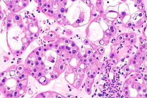
 Hybrid oncocytic/chromophobe tumour of the kidney. H&E stain. | |
|
| |
| Synonyms | hybrid tumour |
|
| |
| LM | features of renal oncocytoma and chromophobe renal cell carcinoma - varies by subtype |
| Subtypes | as per Hes et al.: (1) collision tumour-type, (2) renal oncocytoma with scattered chromophobe cells-type, (3) large eosinophilic cells with intracytoplasmic vacuoles-type |
| LM DDx | renal oncocytoma, chromophobe renal cell carcinoma, renal cell carcinoma, unclassified, SDH-deficient renal cell carcinoma, other renal tumours with eosinophilic cytoplasm |
| Stains | Hale's colloidal iron +ve |
| IHC | CD117 +ve, CK7 +ve (variable) |
| Molecular | no features of ChRCC |
| Grossing notes | partial nephrectomy grossing, total nephrectomy for tumour grossing |
| Site | kidney - see kidney tumours |
|
| |
| Syndromes | Birt–Hogg–Dubé syndrome |
|
| |
| Clinical history | renal mass |
| Prevalence | very rare |
| Prognosis | good on very limited data |
| Treatment | surgical excision |
Renal hybrid oncocytic/chromophobe tumour, also hybrid oncocytic/chromophobe tumour (abbreviated HOCT) and hybrid tumour, is a rare kidney tumour with features of chromophobe renal cell carcinoma and renal oncocytoma.[1]
General
- Rare.
- Molecular heterogeneous group[1] - may represent several different entities.
- Prognosis good - based on one series of 11 cases.[2]
May be seen in several contexts:[1]
- Sporadic.
- Birt–Hogg–Dubé syndrome.
- Renal oncocytosis.
Microscopic
Three morphologic patterns as per Hes et al.:[1]
- Renal oncocytoma and chromophobe renal cell carcinoma collision tumour.
- Different fields viewed in isolation would be compatible with the different diagnoses. Tumour component do not intermingle.
- Renal oncocytoma with scattered chromophobe cells.
- Large eosinophilic cell with intracytoplasmic vacuoles - this is the evolving entity eosinophilic vacuolated tumour.
- Prominent nucleoli (ISUP nucleolar grade 3).
- Perinuclear halos (occasional).
- Nested architecture.
DDx:
- Renal oncocytoma - may have limited chromophobe-like areas (<=5% of tumour).[3]
- Chromophobe renal cell carcinoma, eosinophilic variant.
- SDH-deficient renal cell carcinoma - lower nuclear grade, not nested.
- Eosinophilic vacuolated tumour - evolving entity.
- Other renal tumours with eosinophilic cytoplasm.
- Renal cell carcinoma, unclassified.
Images
Case 1
Case 2
Stains
Features:[2]
- Hale's colloidal iron +ve (apical pattern).
IHC
Features:
Others:
- Vimentin -ve.
- EMA +ve.
- CD10 +ve.
- PAX8 +ve.
Molecular
- No features characteristic of chromophobe RCC on array-CGH analysis.[2]
Sign out
Left Kidney, Partial Nephrectomy:
- Renal tumour with eosinophilic cytoplasm of undetermined malignant potential
  in keeping with the so called "hybrid oncocytic/chromophobe tumour", see comment.
-- Resection margins clear.
-- Tumour limited to kidney.

Comment:
The tumour may be seen in the context of Birt–Hogg–Dubé syndrome.
See also
References
- ↑ 1.0 1.1 1.2 1.3 1.4 Hes, O.; Petersson, F.; Kuroda, N.; Hora, M.; Michal, M. (Oct 2013). "Renal hybrid oncocytic/chromophobe tumors - a review.". Histol Histopathol 28 (10): 1257-64. PMID 23740406.
- ↑ 2.0 2.1 2.2 2.3 Poté, N.; Vieillefond, A.; Couturier, J.; Arrufat, S.; Metzger, I.; Delongchamps, NB.; Camparo, P.; Mège-Lechevallier, F. et al. (Jun 2013). "Hybrid oncocytic/chromophobe renal cell tumours do not display genomic features of chromophobe renal cell carcinomas.". Virchows Arch 462 (6): 633-8. doi:10.1007/s00428-013-1422-4. PMID 23708994.
- ↑ Trpkov, K.; Yilmaz, A.; Uzer, D.; Dishongh, KM.; Quick, CM.; Bismar, TA.; Gokden, N. (Dec 2010). "Renal oncocytoma revisited: a clinicopathological study of 109 cases with emphasis on problematic diagnostic features.". Histopathology 57 (6): 893-906. doi:10.1111/j.1365-2559.2010.03726.x. PMID 21166703.


