Difference between revisions of "Appendix of the epididymis"
Jump to navigation
Jump to search
(→Microscopic: +images) |
|||
| (8 intermediate revisions by the same user not shown) | |||
| Line 1: | Line 1: | ||
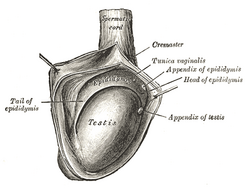
'''Appendix of the epididymis''' is a benign structure derived from the mesonephric duct. | [[Image:Gray1148.png|thumb|right|250px|Drawing showing the appendix of epididymis. (WC/Gray's Anatomy)]] | ||
'''Appendix of the epididymis''', also '''epididymal appendix''', is a benign structure thought to be derived from the mesonephric duct (or Wolffian duct).<ref name=pmid8886956/> | |||
It | It should not be confused with ''[[appendix of the testis]]''. | ||
==General== | |||
*Identifiable on ultrasound.<ref>{{Cite journal | last1 = Kantarci | first1 = F. | last2 = Ozer | first2 = H. | last3 = Adaletli | first3 = I. | last4 = Mihmanli | first4 = I. | title = Cystic appendix epididymis: a sonomorphologic study. | journal = Surg Radiol Anat | volume = 27 | issue = 6 | pages = 557-61 | month = Dec | year = 2005 | doi = 10.1007/s00276-005-0034-3 | PMID = 16195812 }}</ref> | |||
*Seen in approximately 20% of the population.<ref name=pmid8886956>{{Cite journal | last1 = Sahni | first1 = D. | last2 = Jit | first2 = I. | last3 = Joshi | first3 = K. | last4 = Sanjeev | first4 = . | title = Incidence and structure of the appendices of the testis and epididymis. | journal = J Anat | volume = 189 ( Pt 2) | issue = | pages = 341-8 | month = Oct | year = 1996 | doi = | PMID = 8886956 }}</ref> | |||
==Gross== | |||
Features:<ref name=pmid8886956/> | |||
*Thin walled cyst on head of the [[epididymis]]. | |||
*Typically on a stalk. | |||
==Microscopic== | |||
Features:<ref name=pmid8886956/> | |||
*Cyst lined by pseudostratified cilitated epithelium. | |||
*Dense collagen (wall). | |||
*Mesothelium (external aspect). | |||
===Images=== | |||
<gallery> | |||
Image: Appendix of epididymis -- very low mag.jpg | AE - very low mag. | |||
Image: Appendix of epididymis -- low mag.jpg | AE - low mag. | |||
Image: Appendix of epididymis -- intermed mag.jpg | AE - intermed. mag. | |||
Image: Appendix of epididymis -- high mag.jpg | AE - high mag. | |||
Image: Appendix of epididymis -- very high mag.jpg | AE - very hig mag. | |||
Image: Appendix of epididymis - a -- high mag.jpg | AE - high mag. | |||
Image: Appendix of epididymis - a -- very high mag.jpg | AE - very hig mag. | |||
Image: Appendix of epididymis - b -- very high mag.jpg | AE - very hig mag. | |||
</gallery> | |||
==See also== | ==See also== | ||
Latest revision as of 06:57, 22 February 2015
Appendix of the epididymis, also epididymal appendix, is a benign structure thought to be derived from the mesonephric duct (or Wolffian duct).[1]
It should not be confused with appendix of the testis.
General
Gross
Features:[1]
- Thin walled cyst on head of the epididymis.
- Typically on a stalk.
Microscopic
Features:[1]
- Cyst lined by pseudostratified cilitated epithelium.
- Dense collagen (wall).
- Mesothelium (external aspect).
Images
See also
References
- ↑ 1.0 1.1 1.2 1.3 Sahni, D.; Jit, I.; Joshi, K.; Sanjeev, . (Oct 1996). "Incidence and structure of the appendices of the testis and epididymis.". J Anat 189 ( Pt 2): 341-8. PMID 8886956.
- ↑ Kantarci, F.; Ozer, H.; Adaletli, I.; Mihmanli, I. (Dec 2005). "Cystic appendix epididymis: a sonomorphologic study.". Surg Radiol Anat 27 (6): 557-61. doi:10.1007/s00276-005-0034-3. PMID 16195812.


