Difference between revisions of "Uterine cervix with atrophic changes"
Jump to navigation
Jump to search
(+infobox, +images) |
m (touch) |
||
| (5 intermediate revisions by the same user not shown) | |||
| Line 1: | Line 1: | ||
{{ Infobox diagnosis | {{ Infobox diagnosis | ||
| Name = {{PAGENAME}} | | Name = {{PAGENAME}} | ||
| Image = Atrophic cervix -- very high mag.jpg | | Image = Atrophic cervix -- very high mag.jpg | ||
| Width = | | Width = | ||
| Caption = Atrophic cervix. [[H&E stain]]. | | Caption = Atrophic cervix. [[H&E stain]]. | ||
| Line 7: | Line 7: | ||
| Micro = small squamous cells with grey/blue cytoplasm, no "dancing"/"sparkling" chromatin, no mitoses | | Micro = small squamous cells with grey/blue cytoplasm, no "dancing"/"sparkling" chromatin, no mitoses | ||
| Subtypes = | | Subtypes = | ||
| LMDDx = [[HSIL]] | | LMDDx = [[HSIL]], immature [[squamous metaplasia of the uterine cervix|squamous metaplasia]] | ||
| Stains = | | Stains = | ||
| IHC = p16 -ve, Ki-67 rare basal cells | | IHC = p16 -ve, Ki-67 rare basal cells | ||
| Line 18: | Line 18: | ||
| Assdx = | | Assdx = | ||
| Syndromes = | | Syndromes = | ||
| Clinicalhx = | | Clinicalhx = usually postmenopausal | ||
| Signs = | | Signs = | ||
| Symptoms = | | Symptoms = | ||
| Line 40: | Line 40: | ||
==Microscopic== | ==Microscopic== | ||
Features: | Features - squamous cells: | ||
*Cells smaller. | *Cells smaller. | ||
*Cytoplasm grey/blue. | *Cytoplasm grey/blue. | ||
*No "dancing"/"sparkling" chromatin. | *No "dancing"/"sparkling" chromatin. | ||
*No mitoses. | *No mitoses. | ||
Notes: | |||
*Mitosis do not exclude the diagnosis.... but should make one think HSIL. | |||
DDx: | DDx: | ||
*[[HSIL]]. | *[[HSIL]]. | ||
*Immature [[squamous metaplasia of the uterine cervix|squamous metaplasia]].<ref name=pmid17824788/> | |||
===Images=== | ===Images=== | ||
<gallery> | <gallery> | ||
Image: Atrophic cervix -- intermed mag.jpg | AC - intermed. mag. | Image: Atrophic cervix -- intermed mag.jpg | AC - intermed. mag. (WC) | ||
Image: Atrophic cervix - alt -- high mag.jpg | AC - high mag. | Image: Atrophic cervix - alt -- high mag.jpg | AC - high mag. (WC) | ||
Image: Atrophic cervix -- high mag.jpg | AC - high mag. | Image: Atrophic cervix -- high mag.jpg | AC - high mag. (WC) | ||
Image: Atrophic cervix -- very high mag.jpg | AC - very high mag. | Image: Atrophic cervix -- very high mag.jpg | AC - very high mag. (WC) | ||
</gallery> | </gallery> | ||
<gallery> | <gallery> | ||
Image: Atrophic cervix - 2 -- intermed mag.jpg | AC - intermed. mag. | Image: Atrophic cervix - 2 -- intermed mag.jpg | AC - intermed. mag. (WC) | ||
Image: Atrophic cervix - 2 -- high mag.jpg | AC - high mag. | Image: Atrophic cervix - 2 -- high mag.jpg | AC - high mag. (WC) | ||
Image: Atrophic cervix - 2 -- very high mag.jpg | AC - very high mag. | Image: Atrophic cervix - 2 -- very high mag.jpg | AC - very high mag. (WC) | ||
</gallery> | </gallery> | ||
www: | www: | ||
| Line 67: | Line 71: | ||
==IHC== | ==IHC== | ||
Features:<ref name=pmid17824788>{{Cite journal | last1 = Iaconis | first1 = L. | last2 = Hyjek | first2 = E. | last3 = Ellenson | first3 = LH. | last4 = Pirog | first4 = EC. | title = p16 and Ki-67 immunostaining in atypical immature squamous metaplasia of the uterine cervix: correlation with human papillomavirus detection. | journal = Arch Pathol Lab Med | volume = 131 | issue = 9 | pages = 1343-9 | month = Sep | year = 2007 | doi = 10.1043/1543-2165(2007)131[1343:PAKIIA]2.0.CO;2 | PMID = 17824788 }}</ref> | |||
*p16 -ve. | *p16 -ve. | ||
*Ki-67 rare basal cells. | *Ki-67 rare basal cells. | ||
Latest revision as of 13:55, 27 June 2015
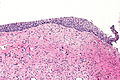
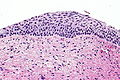
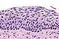
Uterine cervix with atrophic changes is relatively common and is important to recognize as it can mimic HSIL.
| Uterine cervix with atrophic changes | |
|---|---|
| Diagnosis in short | |
 Atrophic cervix. H&E stain. | |
|
| |
| LM | small squamous cells with grey/blue cytoplasm, no "dancing"/"sparkling" chromatin, no mitoses |
| LM DDx | HSIL, immature squamous metaplasia |
| IHC | p16 -ve, Ki-67 rare basal cells |
| Site | uterine cervix - exocervix |
|
| |
| Clinical history | usually postmenopausal |
| Prevalence | common |
| Prognosis | benign |
| Other | normal - postmenopausal |
It is also known as atrophy of the uterine cervix, cervical atrophy, atrophy of the cervix and cervix with atrophic changes.
General
- Common.
- Post-menupausal.
- Important to recognize and differentiate from HSIL.
Microscopic
Features - squamous cells:
- Cells smaller.
- Cytoplasm grey/blue.
- No "dancing"/"sparkling" chromatin.
- No mitoses.
Notes:
- Mitosis do not exclude the diagnosis.... but should make one think HSIL.
DDx:
- HSIL.
- Immature squamous metaplasia.[1]
Images
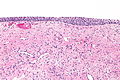
AC - intermed. mag. (WC)
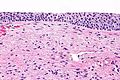
AC - high mag. (WC)
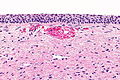
AC - high mag. (WC)
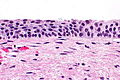
AC - very high mag. (WC)


AC - intermed. mag. (WC)
AC - high mag. (WC)
AC - very high mag. (WC)


www:
IHC
Features:[1]
- p16 -ve.
- Ki-67 rare basal cells.
Sign out
UTERINE CERVIX, BIOPSY:
- SQUAMOUS MUCOSA WITH ATROPHIC CHANGES.
- BENIGN ENDOCERVICAL EPITHELIUM.
- NEGATIVE FOR DYSPLASIA.

COMMENT:
A p16 immunostain is negative. A Ki-67 immunostain marks rare basal cells.
See also
References
- ↑ 1.0 1.1 Iaconis, L.; Hyjek, E.; Ellenson, LH.; Pirog, EC. (Sep 2007). "p16 and Ki-67 immunostaining in atypical immature squamous metaplasia of the uterine cervix: correlation with human papillomavirus detection.". Arch Pathol Lab Med 131 (9): 1343-9. doi:10.1043/1543-2165(2007)131[1343:PAKIIA]2.0.CO;2. PMID 17824788.
- ↑ URL: http://www.eurocytology.eu/static/eurocytology/TUR/cervical/LP1ContentLcontC.html. Accessed on: 13 December 2013.