Difference between revisions of "Suture material"
Jump to navigation
Jump to search
(Created page with "'''Suture material''' is occasionally seen under the microscope. It is usually easy to identified and typically polarizes. ===Images=== <gallery> Image:Solitary_fibrous_t...") |
Alessandro (talk | contribs) m (→Microscopic) |
||
| (8 intermediate revisions by one other user not shown) | |||
| Line 1: | Line 1: | ||
'''Suture material''' is occasionally seen under the [[microscope]]. It is usually easy to identified and typically polarizes. | '''Suture material''' is occasionally seen under the [[microscope]]. It is usually easy to identified and typically polarizes. | ||
==General== | |||
*Suture are often used to orient a specimen<ref name=pmid23287819>{{Cite journal | last1 = Volleamere | first1 = AJ. | last2 = Kirwan | first2 = CC. | title = National survey of breast cancer specimen orientation marking systems. | journal = Eur J Surg Oncol | volume = 39 | issue = 3 | pages = 255-9 | month = Mar | year = 2013 | doi = 10.1016/j.ejso.2012.12.008 | PMID = 23287819 }}</ref> and/or mark the true [[surgical margin]].<ref name=pmid7544556>{{Cite journal | last1 = Seitz | first1 = SE. | last2 = Foley | first2 = GL. | last3 = Marretta | first3 = SM. | title = Evaluation of marking materials for cutaneous surgical margins. | journal = Am J Vet Res | volume = 56 | issue = 6 | pages = 826-33 | month = Jun | year = 1995 | doi = | PMID = 7544556 }}</ref> | |||
==Microscopic== | |||
Features: | |||
*Glassy appearance - sharply circumscribed. | |||
*+/-Tearing surrounding tissue. | |||
*+/-Foreign body-type [[granuloma]]s with multinucleated giant cells. | |||
**Seen only if the suture has been in place for at least several days. | |||
===Images=== | ===Images=== | ||
<gallery> | <gallery> | ||
Image:Solitary_fibrous_tumour_low_mag.jpg | Suture material adjacent to a [[solitary fibrous tumour|SFT]]. | Image:Solitary_fibrous_tumour_low_mag.jpg | Suture material adjacent to a [[solitary fibrous tumour|SFT]]. | ||
Image:Suture_micrograph.jpg | Suture material. | |||
</gallery> | </gallery> | ||
==See also== | ==See also== | ||
*[[Histology artifacts]]. | *[[Histology artifacts]]. | ||
*[[Foreign material]]. | |||
*[[Granuloma]]. | |||
==References== | |||
{{Reflist|2}} | |||
[[Category:Basics]] | [[Category:Basics]] | ||
Latest revision as of 13:17, 11 February 2019
Suture material is occasionally seen under the microscope. It is usually easy to identified and typically polarizes.
General
- Suture are often used to orient a specimen[1] and/or mark the true surgical margin.[2]
Microscopic
Features:
- Glassy appearance - sharply circumscribed.
- +/-Tearing surrounding tissue.
- +/-Foreign body-type granulomas with multinucleated giant cells.
- Seen only if the suture has been in place for at least several days.
Images
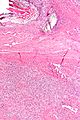
Suture material adjacent to a SFT.
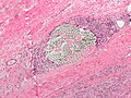
Suture material.


See also
References
- ↑ Volleamere, AJ.; Kirwan, CC. (Mar 2013). "National survey of breast cancer specimen orientation marking systems.". Eur J Surg Oncol 39 (3): 255-9. doi:10.1016/j.ejso.2012.12.008. PMID 23287819.
- ↑ Seitz, SE.; Foley, GL.; Marretta, SM. (Jun 1995). "Evaluation of marking materials for cutaneous surgical margins.". Am J Vet Res 56 (6): 826-33. PMID 7544556.