Difference between revisions of "Xanthogranulomatous cholecystitis"
Jump to navigation
Jump to search
(tweak) |
|||
| (15 intermediate revisions by the same user not shown) | |||
| Line 1: | Line 1: | ||
'''Xanthogranulomatous cholecystitis''', abbreviated '''XGC''', is an uncommon pathology of the [[gallbladder]]. | {{ Infobox diagnosis | ||
| Name = {{PAGENAME}} | |||
| Image = Xanthogranulomatous cholecystitis -- low mag.jpg | |||
| Width = | |||
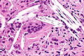
| Caption = Xanthogranulomatous cholecystitis. [[H&E stain]]. | |||
| Micro = cholesterol clefts, [[granulomas]] | |||
| Subtypes = | |||
| LMDDx = [[chronic cholecystitis]], [[gallbladder cholesterolosis]] | |||
| Stains = | |||
| IHC = | |||
| EM = | |||
| Molecular = | |||
| IF = | |||
| Gross = thickened gallbladder wall, [[gallstones]] | |||
| Grossing = | |||
| Site = [[gallbladder]] | |||
| Assdx = | |||
| Syndromes = | |||
| Clinicalhx = | |||
| Signs = | |||
| Symptoms = | |||
| Prevalence = uncommon | |||
| Bloodwork = | |||
| Rads = hypo-attenuated nodules in gallbladder wall | |||
| Endoscopy = | |||
| Prognosis = benign | |||
| Other = | |||
| ClinDDx = [[gallbladder carcinoma]], [[acute cholecystitis]] | |||
| Tx = cholecystectomy (surgical removal) | |||
}} | |||
'''Xanthogranulomatous cholecystitis''', abbreviated '''XGC''',<ref name=pmid25404941>{{Cite journal | last1 = Rammohan | first1 = A. | last2 = Cherukuri | first2 = SD. | last3 = Sathyanesan | first3 = J. | last4 = Palaniappan | first4 = R. | last5 = Govindan | first5 = M. | title = Xanthogranulomatous cholecystitis masquerading as gallbladder cancer: can it be diagnosed preoperatively? | journal = Gastroenterol Res Pract | volume = 2014 | issue = | pages = 253645 | month = | year = 2014 | doi = 10.1155/2014/253645 | PMID = 25404941 }}</ref> is an uncommon pathology of the [[gallbladder]]. | |||
==General== | ==General== | ||
*Uncommon ~ 1-9%.<ref name=pmid23991684>{{Cite journal | last1 = Hale | first1 = MD. | last2 = Roberts | first2 = KJ. | last3 = Hodson | first3 = J. | last4 = Scott | first4 = N. | last5 = Sheridan | first5 = M. | last6 = Toogood | first6 = GJ. | title = Xanthogranulomatous cholecystitis: a European and global perspective. | journal = HPB (Oxford) | volume = | issue = | pages = | month = Aug | year = 2013 | doi = 10.1111/hpb.12152 | PMID = 23991684 }}</ref><ref name=pmid24019688>{{Cite journal | last1 = Alvi | first1 = AR. | last2 = Jalbani | first2 = I. | last3 = Murtaza | first3 = G. | last4 = Hameed | first4 = A. | title = Outcomes of Xanthogranulomatous cholecystitis in laparoscopic era: A retrospective Cohort study. | journal = J Minim Access Surg | volume = 9 | issue = 3 | pages = 109-15 | month = Jul | year = 2013 | doi = 10.4103/0972-9941.115368 | PMID = 24019688 }}</ref> | *Uncommon ~ 1-9%.<ref name=pmid23991684>{{Cite journal | last1 = Hale | first1 = MD. | last2 = Roberts | first2 = KJ. | last3 = Hodson | first3 = J. | last4 = Scott | first4 = N. | last5 = Sheridan | first5 = M. | last6 = Toogood | first6 = GJ. | title = Xanthogranulomatous cholecystitis: a European and global perspective. | journal = HPB (Oxford) | volume = | issue = | pages = | month = Aug | year = 2013 | doi = 10.1111/hpb.12152 | PMID = 23991684 }}</ref><ref name=pmid24019688>{{Cite journal | last1 = Alvi | first1 = AR. | last2 = Jalbani | first2 = I. | last3 = Murtaza | first3 = G. | last4 = Hameed | first4 = A. | title = Outcomes of Xanthogranulomatous cholecystitis in laparoscopic era: A retrospective Cohort study. | journal = J Minim Access Surg | volume = 9 | issue = 3 | pages = 109-15 | month = Jul | year = 2013 | doi = 10.4103/0972-9941.115368 | PMID = 24019688 }}</ref> | ||
*May be confused with [[gallbladder carcinoma]].<ref name=pmid23991684/> | **Approximately 2% in one series of 724 gallbladders.<ref name=pmid3584484>{{Cite journal | last1 = Roberts | first1 = KM. | last2 = Parsons | first2 = MA. | title = Xanthogranulomatous cholecystitis: clinicopathological study of 13 cases. | journal = J Clin Pathol | volume = 40 | issue = 4 | pages = 412-7 | month = Apr | year = 1987 | doi = | PMID = 3584484 | PMC = 1140974 }}</ref> | ||
*May be confused (clinically) with [[gallbladder carcinoma]].<ref name=pmid25404941/><ref name=pmid23991684/><ref name=pmid23060404>{{Cite journal | last1 = Martins | first1 = PN. | last2 = Sheiner | first2 = P. | last3 = Facciuto | first3 = M. | title = Xanthogranulomatous cholecystitis mimicking gallbladder cancer and causing obstructive cholestasis. | journal = Hepatobiliary Pancreat Dis Int | volume = 11 | issue = 5 | pages = 549-52 | month = Oct | year = 2012 | doi = | PMID = 23060404 }}</ref> | |||
==Gross== | ==Gross== | ||
| Line 15: | Line 46: | ||
==Microscopic== | ==Microscopic== | ||
Features: | Features:<ref name=pmid3584484>{{Cite journal | last1 = Roberts | first1 = KM. | last2 = Parsons | first2 = MA. | title = Xanthogranulomatous cholecystitis: clinicopathological study of 13 cases. | journal = J Clin Pathol | volume = 40 | issue = 4 | pages = 412-7 | month = Apr | year = 1987 | doi = | PMID = 3584484 | PMC = 1140974 }}</ref> | ||
*[[Granulomas]]. | *[[Granulomas]]. | ||
*Lipid-laden macrophages. | |||
*+/-Cholesterol clefts. | |||
*Inflammatory cells. | |||
*Fibrosis. | |||
DDx: | |||
*[[Chronic cholecystitis]]. | |||
*[[Gallbladder cholesterolosis]]. | |||
===Images=== | |||
<gallery> | |||
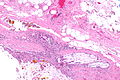
Image: Xanthogranulomatous cholecystitis -- low mag.jpg | XGC - low mag. (WC) | |||
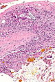
Image: Xanthogranulomatous cholecystitis -- intermed mag.jpg | XGC - intermed. mag. (WC) | |||
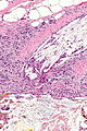
Image: Xanthogranulomatous cholecystitis - alt -- intermed mag.jpg | XGC - intermed. mag. (WC) | |||
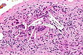
Image: Xanthogranulomatous cholecystitis -- high mag.jpg | XGC - high mag. (WC) | |||
Image: Xanthogranulomatous cholecystitis -- very high mag.jpg | XGC - very high mag. (WC) | |||
</gallery> | |||
www: | |||
*[http://www.radiologycases.com/index.php/radiologycases/article/view/696 XGC (radiologycases.com)].<ref>{{Cite journal | last1 = Cecava | first1 = ND. | last2 = Andrews | first2 = R. | title = Case report of xanthogranulomatous cholecystitis, review of its sonographic and magnetic resonance findings, and distinction from other gallbladder pathology. | journal = J Radiol Case Rep | volume = 5 | issue = 4 | pages = 19-24 | month = | year = 2011 | doi = 10.3941/jrcr.v5i4.696 | PMID = 22470787 }}</ref> | |||
*[http://www.ncbi.nlm.nih.gov/pmc/articles/PMC4227324/figure/fig3/ XGC (nih.gov)].<ref name=pmid25404941/> | |||
==Sign out== | |||
<pre> | |||
Gallbladder, Cholecystectomy: | |||
- Xanthogranulomatous cholecystitis. | |||
- Cholelithiasis. | |||
</pre> | |||
===Block letters=== | |||
<pre> | |||
GALLBLADDER, CHOLECYSTECTOMY: | |||
- XANTHOGRANULOMATOUS CHOLECYSTITIS. | |||
- CHOLELITHIASIS. | |||
</pre> | |||
===Micro=== | |||
The sections show a thickened gallbladder wall with cholesterol clefts, multinucleated | |||
giant cells, fibrosis and lymphoid aggregates. No metaplasia, nuclear atypia or dysplasia | |||
is apparent. | |||
====Alternate==== | |||
The sections show a thickened gallbladder wall with cholesterol clefts, multinucleated | |||
giant cells, fibrosis and small lymphoid aggregates. No metaplasia, dysplasia or | |||
significant nuclear atypia is apparent. | |||
==See also== | ==See also== | ||
Latest revision as of 03:54, 31 May 2016
| Xanthogranulomatous cholecystitis | |
|---|---|
| Diagnosis in short | |
 Xanthogranulomatous cholecystitis. H&E stain. | |
|
| |
| LM | cholesterol clefts, granulomas |
| LM DDx | chronic cholecystitis, gallbladder cholesterolosis |
| Gross | thickened gallbladder wall, gallstones |
| Site | gallbladder |
|
| |
| Prevalence | uncommon |
| Radiology | hypo-attenuated nodules in gallbladder wall |
| Prognosis | benign |
| Clin. DDx | gallbladder carcinoma, acute cholecystitis |
| Treatment | cholecystectomy (surgical removal) |
Xanthogranulomatous cholecystitis, abbreviated XGC,[1] is an uncommon pathology of the gallbladder.
General
- Uncommon ~ 1-9%.[2][3]
- Approximately 2% in one series of 724 gallbladders.[4]
- May be confused (clinically) with gallbladder carcinoma.[1][2][5]
Gross
Features:[6]
- Wall thickening ~ 90% of cases.
- Gallstones ~ 70% of cases.
- +/-Infiltration of surrounding tissues (liver, fat).
Imaging:
- Hypo-attenuated nodules in the gallbladder wall.[6]
Microscopic
Features:[4]
- Granulomas.
- Lipid-laden macrophages.
- +/-Cholesterol clefts.
- Inflammatory cells.
- Fibrosis.
DDx:
Images
XGC - low mag. (WC)
XGC - intermed. mag. (WC)
XGC - intermed. mag. (WC)
XGC - high mag. (WC)
XGC - very high mag. (WC)


www:
Sign out
Gallbladder, Cholecystectomy:
- Xanthogranulomatous cholecystitis.
- Cholelithiasis.
Block letters
GALLBLADDER, CHOLECYSTECTOMY:
- XANTHOGRANULOMATOUS CHOLECYSTITIS.
- CHOLELITHIASIS.
Micro
The sections show a thickened gallbladder wall with cholesterol clefts, multinucleated giant cells, fibrosis and lymphoid aggregates. No metaplasia, nuclear atypia or dysplasia is apparent.
Alternate
The sections show a thickened gallbladder wall with cholesterol clefts, multinucleated giant cells, fibrosis and small lymphoid aggregates. No metaplasia, dysplasia or significant nuclear atypia is apparent.
See also
References
- ↑ 1.0 1.1 1.2 Rammohan, A.; Cherukuri, SD.; Sathyanesan, J.; Palaniappan, R.; Govindan, M. (2014). "Xanthogranulomatous cholecystitis masquerading as gallbladder cancer: can it be diagnosed preoperatively?". Gastroenterol Res Pract 2014: 253645. doi:10.1155/2014/253645. PMID 25404941.
- ↑ 2.0 2.1 Hale, MD.; Roberts, KJ.; Hodson, J.; Scott, N.; Sheridan, M.; Toogood, GJ. (Aug 2013). "Xanthogranulomatous cholecystitis: a European and global perspective.". HPB (Oxford). doi:10.1111/hpb.12152. PMID 23991684.
- ↑ Alvi, AR.; Jalbani, I.; Murtaza, G.; Hameed, A. (Jul 2013). "Outcomes of Xanthogranulomatous cholecystitis in laparoscopic era: A retrospective Cohort study.". J Minim Access Surg 9 (3): 109-15. doi:10.4103/0972-9941.115368. PMID 24019688.
- ↑ 4.0 4.1 Roberts, KM.; Parsons, MA. (Apr 1987). "Xanthogranulomatous cholecystitis: clinicopathological study of 13 cases.". J Clin Pathol 40 (4): 412-7. PMC 1140974. PMID 3584484. https://www.ncbi.nlm.nih.gov/pmc/articles/PMC1140974/.
- ↑ Martins, PN.; Sheiner, P.; Facciuto, M. (Oct 2012). "Xanthogranulomatous cholecystitis mimicking gallbladder cancer and causing obstructive cholestasis.". Hepatobiliary Pancreat Dis Int 11 (5): 549-52. PMID 23060404.
- ↑ 6.0 6.1 Zhao, F.; Lu, PX.; Yan, SX.; Wang, GF.; Yuan, J.; Zhang, SZ.; Wang, YX. (Sep 2013). "CT and MR features of xanthogranulomatous cholecystitis: an analysis of consecutive 49 cases.". Eur J Radiol 82 (9): 1391-7. doi:10.1016/j.ejrad.2013.04.026. PMID 23726123.
- ↑ Cecava, ND.; Andrews, R. (2011). "Case report of xanthogranulomatous cholecystitis, review of its sonographic and magnetic resonance findings, and distinction from other gallbladder pathology.". J Radiol Case Rep 5 (4): 19-24. doi:10.3941/jrcr.v5i4.696. PMID 22470787.