Difference between revisions of "Leukoplakia"
Jump to navigation
Jump to search
(redirect) |
|||
| (3 intermediate revisions by the same user not shown) | |||
| Line 1: | Line 1: | ||
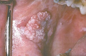
[[Image:Leucoexo.jpg|thumb|right|Leukoplakia. (WC/Aitor III)]] | |||
'''Leukoplakia''' is a relatively common clinical finding in clinical medicine. This article looks at leukoplakia of the [[head and neck pathology|head and neck]]. | |||
''[[Hairy leukoplakia]]'' is dealt with separately. The typical ''[[benign leukoplakia]]'' is also dealt with separately. | |||
==General== | |||
*Non-specific clinical finding - may be benign ''or'' [[malignant]]. | |||
*Associated with [[tobacco]] use.<ref name=pmid11336117>{{Cite journal | last1 = Bánóczy | first1 = J. | last2 = Gintner | first2 = Z. | last3 = Dombi | first3 = C. | title = Tobacco use and oral leukoplakia. | journal = J Dent Educ | volume = 65 | issue = 4 | pages = 322-7 | month = Apr | year = 2001 | doi = | PMID = 11336117 }}</ref> | |||
Risk of malignancy: | |||
*In twos series ~13% were associated with an invasive lesion.<ref name=pmid19953947>{{Cite journal | last1 = Lan | first1 = AX. | last2 = Guan | first2 = XB. | last3 = Sun | first3 = Z. | title = [Analysis of risk factors for carcinogenesis of oral leukoplakia]. | journal = Zhonghua Kou Qiang Yi Xue Za Zhi | volume = 44 | issue = 6 | pages = 327-31 | month = Jun | year = 2009 | doi = | PMID = 19953947 }}</ref><ref name=pmid16545712>{{Cite journal | last1 = Lee | first1 = JJ. | last2 = Hung | first2 = HC. | last3 = Cheng | first3 = SJ. | last4 = Chen | first4 = YJ. | last5 = Chiang | first5 = CP. | last6 = Liu | first6 = BY. | last7 = Jeng | first7 = JH. | last8 = Chang | first8 = HH. | last9 = Kuo | first9 = YS. | title = Carcinoma and dysplasia in oral leukoplakias in Taiwan: prevalence and risk factors. | journal = Oral Surg Oral Med Oral Pathol Oral Radiol Endod | volume = 101 | issue = 4 | pages = 472-80 | month = Apr | year = 2006 | doi = 10.1016/j.tripleo.2005.07.024 | PMID = 16545712 }}</ref> | |||
*Non-homogenous leukoplakia has a greater risk of malignancy than homogenous.<ref name=pmid16545712/> | |||
*Location matters - floor of mouth and ventral tongue lesions higher risk for malignancy.<ref name=pmid7548621>{{Cite journal | last1 = Sciubba | first1 = JJ. | title = Oral leukoplakia. | journal = Crit Rev Oral Biol Med | volume = 6 | issue = 2 | pages = 147-60 | month = | year = 1995 | doi = | PMID = 7548621 | URL = http://cro.sagepub.com/content/6/2/147.long }}</ref> | |||
==Gross== | |||
*White lesion - may be subdivided: | |||
**Non-homogenous. | |||
**Homogenous. | |||
==Microscopic== | |||
Features:<ref name=Ref_PBoD780>{{Ref PBoD|780}}</ref> | |||
*Often associated with epithelial thickening ([[hyperkeratosis]], [[acanthosis]]). | |||
DDx: | |||
*Food debris. | |||
*[[Oral candidiasis]]. | |||
*[[Lichen planus]]. | |||
*[[Benign alveolar ridge keratosis]] (oral [[lichen simplex chronicus]]).<ref name=pmid18158926>{{Cite journal | last1 = Natarajan | first1 = E. | last2 = Woo | first2 = SB. | title = Benign alveolar ridge keratosis (oral lichen simplex chronicus): A distinct clinicopathologic entity. | journal = J Am Acad Dermatol | volume = 58 | issue = 1 | pages = 151-7 | month = Jan | year = 2008 | doi = 10.1016/j.jaad.2007.07.011 | PMID = 18158926 }}</ref> | |||
*[[Squamous cell carcinoma of the head and neck]]. | |||
*Others - see ''[[Dermatopathology#Leukoplakia]]''. | |||
==See also== | |||
*[[An introduction to head and neck pathology]]. | |||
*[[Erythroplakia]]. | |||
*[[Leukoedema]]. | |||
==References== | |||
{{Reflist|2}} | |||
[[Category:Clinical]] | |||
[[Category:Head and neck pathology]] | |||
Latest revision as of 18:16, 2 May 2017
Leukoplakia is a relatively common clinical finding in clinical medicine. This article looks at leukoplakia of the head and neck.
Hairy leukoplakia is dealt with separately. The typical benign leukoplakia is also dealt with separately.
General
Risk of malignancy:
- In twos series ~13% were associated with an invasive lesion.[2][3]
- Non-homogenous leukoplakia has a greater risk of malignancy than homogenous.[3]
- Location matters - floor of mouth and ventral tongue lesions higher risk for malignancy.[4]
Gross
- White lesion - may be subdivided:
- Non-homogenous.
- Homogenous.
Microscopic
Features:[5]
- Often associated with epithelial thickening (hyperkeratosis, acanthosis).
DDx:
- Food debris.
- Oral candidiasis.
- Lichen planus.
- Benign alveolar ridge keratosis (oral lichen simplex chronicus).[6]
- Squamous cell carcinoma of the head and neck.
- Others - see Dermatopathology#Leukoplakia.
See also
References
- ↑ Bánóczy, J.; Gintner, Z.; Dombi, C. (Apr 2001). "Tobacco use and oral leukoplakia.". J Dent Educ 65 (4): 322-7. PMID 11336117.
- ↑ Lan, AX.; Guan, XB.; Sun, Z. (Jun 2009). "[Analysis of risk factors for carcinogenesis of oral leukoplakia].". Zhonghua Kou Qiang Yi Xue Za Zhi 44 (6): 327-31. PMID 19953947.
- ↑ 3.0 3.1 Lee, JJ.; Hung, HC.; Cheng, SJ.; Chen, YJ.; Chiang, CP.; Liu, BY.; Jeng, JH.; Chang, HH. et al. (Apr 2006). "Carcinoma and dysplasia in oral leukoplakias in Taiwan: prevalence and risk factors.". Oral Surg Oral Med Oral Pathol Oral Radiol Endod 101 (4): 472-80. doi:10.1016/j.tripleo.2005.07.024. PMID 16545712.
- ↑ Sciubba, JJ. (1995). "Oral leukoplakia.". Crit Rev Oral Biol Med 6 (2): 147-60. PMID 7548621.
- ↑ Cotran, Ramzi S.; Kumar, Vinay; Fausto, Nelson; Nelso Fausto; Robbins, Stanley L.; Abbas, Abul K. (2005). Robbins and Cotran pathologic basis of disease (7th ed.). St. Louis, Mo: Elsevier Saunders. pp. 780. ISBN 0-7216-0187-1.
- ↑ Natarajan, E.; Woo, SB. (Jan 2008). "Benign alveolar ridge keratosis (oral lichen simplex chronicus): A distinct clinicopathologic entity.". J Am Acad Dermatol 58 (1): 151-7. doi:10.1016/j.jaad.2007.07.011. PMID 18158926.
