Difference between revisions of "Cystitis cystica"
Jump to navigation
Jump to search
(redirect) |
|||
| (8 intermediate revisions by the same user not shown) | |||
| Line 1: | Line 1: | ||
{{ Infobox diagnosis | |||
| Name = {{PAGENAME}} | |||
| Image = Cystitis_cystica_-_alt_--_intermed_mag.jpg | |||
| Width = | |||
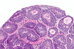
| Caption = Cystitis cystica. [[H&E stain]]. | |||
| Synonyms = | |||
| Micro = nests of urothelium within the lamina propria with cyst formation, i.e. lumens are present; no nuclear atypia | |||
| Subtypes = | |||
| LMDDx = [[nested urothelial carcinoma]], [[cystitis cystica et glandularis]], [[inverted urothelial papilloma]] | |||
| Stains = | |||
| IHC = | |||
| EM = | |||
| Molecular = | |||
| IF = | |||
| Gross = | |||
| Grossing = | |||
| Site = [[urinary bladder]] - see ''[[urothelium]]'' | |||
| Assdx = | |||
| Syndromes = | |||
| Clinicalhx = | |||
| Signs = | |||
| Symptoms = | |||
| Prevalence = common | |||
| Bloodwork = | |||
| Rads = | |||
| Endoscopy = | |||
| Prognosis = benign | |||
| Other = | |||
| ClinDDx = [[urothelial carcinoma]] | |||
| Tx = | |||
}} | |||
'''Cystitis cystica''' is a common inflammatory process of the [[urinary bladder]] with cyst formation. | |||
==General== | |||
*Benign. | |||
*Can be thought of as [[von Brunn nests]] with cystic change.<ref name=Ref_WMSP304>{{Ref WMSP|304}}</ref> | |||
*Called ''[[ureteritis cystica]]'' if it happens in a [[ureter]].<ref name=pmid21966620 >{{Cite journal | last1 = Rothschild | first1 = JG. | last2 = Wu | first2 = G. | title = Ureteritis cystica: a radiologic pathologic correlation. | journal = J Clin Imaging Sci | volume = 1 | issue = | pages = 23 | month = | year = 2011 | doi = 10.4103/2156-7514.80375 | PMID = 21966620 }}</ref> | |||
**There is also a ''urethritis cystica'' - seen in the [[urethra]].<ref name=pmid22397870>{{Cite journal | last1 = Conces | first1 = MR. | last2 = Williamson | first2 = SR. | last3 = Montironi | first3 = R. | last4 = Lopez-Beltran | first4 = A. | last5 = Scarpelli | first5 = M. | last6 = Cheng | first6 = L. | title = Urethral caruncle: clinicopathologic features of 41 cases. | journal = Hum Pathol | volume = 43 | issue = 9 | pages = 1400-4 | month = Sep | year = 2012 | doi = 10.1016/j.humpath.2011.10.015 | PMID = 22397870 }}</ref> | |||
==Microscopic== | |||
Features:<ref name=Ref_PBoD1028>{{Ref PBoD|1028}}</ref> | |||
*Nests of urothelium within the lamina propria with cyst formation, i.e. lumens are present. | |||
Note: | |||
*Nests should '''not''' extend into the muscularis propria. | |||
DDx: | |||
*[[Nested urothelial carcinoma]].<ref name=pmid19800100>{{Cite journal | last1 = Wasco | first1 = MJ. | last2 = Daignault | first2 = S. | last3 = Bradley | first3 = D. | last4 = Shah | first4 = RB. | title = Nested variant of urothelial carcinoma: a clinicopathologic and immunohistochemical study of 30 pure and mixed cases. | journal = Hum Pathol | volume = 41 | issue = 2 | pages = 163-71 | month = Feb | year = 2010 | doi = 10.1016/j.humpath.2009.07.015 | PMID = 19800100 }} | |||
</ref> | |||
*[[Cystitis cystica et glandularis]] - has goblet cells. | |||
*[[Inverted urothelial papilloma]] - peripheral palisading, higher architectural complexity. | |||
===Images=== | |||
<gallery> | |||
Image: Cystitis cystica -- low mag.jpg | CC - low mag. | |||
Image: Cystitis cystica -- intermed mag.jpg | CC - intermed. mag. | |||
Image: Cystitis cystica - rot -- intermed mag.jpg | CC - intermed. mag. | |||
Image: Cystitis cystica -- high mag.jpg | CC - high mag. | |||
Image: Cystitis cystica - alt -- intermed mag.jpg | CC - intermed. mag. | |||
Image: Cystitis cystica - alt -- high mag.jpg | CC - high mag. | |||
</gallery> | |||
www: | |||
*[http://www.webpathology.com/image.asp?n=1&Case=50 Cystitis cystica (webpathology.com)]. | |||
==Sign out== | |||
<pre> | |||
URINARY BLADDER, BIOPSY: | |||
- CYSTITIS CYSTICA. | |||
- NEGATIVE FOR MALIGNANCY. | |||
</pre> | |||
<pre> | |||
URINARY BLADDER, TRANSURETHRAL RESECTION: | |||
- CYSTITIS CYSTICA ET GLANDULARIS, FOCALLY WITH LARGE THIN-WALLED CYSTS. | |||
- PROMINENT BENIGN DILATED SUPERFICIAL BLOOD VESSELS, FOCAL. | |||
- NEGATIVE FOR MALIGNANCY. | |||
</pre> | |||
<pre> | |||
URINARY BLADDER (TRIGONE), TRANSURETHRAL BIOPSY: | |||
- SMALL POLYPOID FRAGMENT OF UROTHELIUM CONSISTENT WITH CYSTITIS CYSTICA. | |||
- NO SQUAMOUS METAPLASTIC CHANGE IDENTIFIED. | |||
- NO EVIDENCE OF MALIGNANCY. | |||
</pre> | |||
===Micro=== | |||
The sections show a small fragment of urothelial mucosa with small urothelial nests that are focally cystic. The nests are well-circumscribed and approximately of equal size. Adjacent to the nests is a small cluster of bland appearing lymphocytes. | |||
No exophytic component is identified. No papillary structures are apparent. The cytomorphology of the urothelium is bland and no mitotic activity is readily apparent. | |||
==See also== | |||
*[[Urothelium]]. | |||
*[[Cystitis glandularis]]. | |||
==References== | |||
{{Reflist|2}} | |||
[[Category:Diagnosis]] | [[Category:Diagnosis]] | ||
[[Category:Urothelium]] | |||
Latest revision as of 12:59, 2 November 2018
| Cystitis cystica | |
|---|---|
| Diagnosis in short | |
 Cystitis cystica. H&E stain. | |
|
| |
| LM | nests of urothelium within the lamina propria with cyst formation, i.e. lumens are present; no nuclear atypia |
| LM DDx | nested urothelial carcinoma, cystitis cystica et glandularis, inverted urothelial papilloma |
| Site | urinary bladder - see urothelium |
|
| |
| Prevalence | common |
| Prognosis | benign |
| Clin. DDx | urothelial carcinoma |
Cystitis cystica is a common inflammatory process of the urinary bladder with cyst formation.
General
- Benign.
- Can be thought of as von Brunn nests with cystic change.[1]
- Called ureteritis cystica if it happens in a ureter.[2]
Microscopic
Features:[4]
- Nests of urothelium within the lamina propria with cyst formation, i.e. lumens are present.
Note:
- Nests should not extend into the muscularis propria.
DDx:
- Nested urothelial carcinoma.[5]
- Cystitis cystica et glandularis - has goblet cells.
- Inverted urothelial papilloma - peripheral palisading, higher architectural complexity.
Images
www:
Sign out
URINARY BLADDER, BIOPSY:
- CYSTITIS CYSTICA.
- NEGATIVE FOR MALIGNANCY.
URINARY BLADDER, TRANSURETHRAL RESECTION:
- CYSTITIS CYSTICA ET GLANDULARIS, FOCALLY WITH LARGE THIN-WALLED CYSTS.
- PROMINENT BENIGN DILATED SUPERFICIAL BLOOD VESSELS, FOCAL.
- NEGATIVE FOR MALIGNANCY.
URINARY BLADDER (TRIGONE), TRANSURETHRAL BIOPSY:
- SMALL POLYPOID FRAGMENT OF UROTHELIUM CONSISTENT WITH CYSTITIS CYSTICA.
- NO SQUAMOUS METAPLASTIC CHANGE IDENTIFIED.
- NO EVIDENCE OF MALIGNANCY.
Micro
The sections show a small fragment of urothelial mucosa with small urothelial nests that are focally cystic. The nests are well-circumscribed and approximately of equal size. Adjacent to the nests is a small cluster of bland appearing lymphocytes.
No exophytic component is identified. No papillary structures are apparent. The cytomorphology of the urothelium is bland and no mitotic activity is readily apparent.
See also
References
- ↑ Humphrey, Peter A; Dehner, Louis P; Pfeifer, John D (2008). The Washington Manual of Surgical Pathology (1st ed.). Lippincott Williams & Wilkins. pp. 304. ISBN 978-0781765275.
- ↑ Rothschild, JG.; Wu, G. (2011). "Ureteritis cystica: a radiologic pathologic correlation.". J Clin Imaging Sci 1: 23. doi:10.4103/2156-7514.80375. PMID 21966620.
- ↑ Conces, MR.; Williamson, SR.; Montironi, R.; Lopez-Beltran, A.; Scarpelli, M.; Cheng, L. (Sep 2012). "Urethral caruncle: clinicopathologic features of 41 cases.". Hum Pathol 43 (9): 1400-4. doi:10.1016/j.humpath.2011.10.015. PMID 22397870.
- ↑ Cotran, Ramzi S.; Kumar, Vinay; Fausto, Nelson; Nelso Fausto; Robbins, Stanley L.; Abbas, Abul K. (2005). Robbins and Cotran pathologic basis of disease (7th ed.). St. Louis, Mo: Elsevier Saunders. pp. 1028. ISBN 0-7216-0187-1.
- ↑ Wasco, MJ.; Daignault, S.; Bradley, D.; Shah, RB. (Feb 2010). "Nested variant of urothelial carcinoma: a clinicopathologic and immunohistochemical study of 30 pure and mixed cases.". Hum Pathol 41 (2): 163-71. doi:10.1016/j.humpath.2009.07.015. PMID 19800100.


