Difference between revisions of "Ovarian microcystic stromal tumour"
Jump to navigation
Jump to search
| (4 intermediate revisions by the same user not shown) | |||
| Line 1: | Line 1: | ||
{{ Infobox diagnosis | {{ Infobox diagnosis | ||
| Name = {{PAGENAME}} | | Name = {{PAGENAME}} | ||
| Image = | | Image = Microcystic stromal tumour of ovary -- intermed mag.jpg | ||
| Width = | | Width = | ||
| Caption = | | Caption = Microcystic stromal tumour of ovary. [[H&E stain]]. | ||
| Synonyms = | | Synonyms = | ||
| Micro = microscopic cysts alternating with solid cellular regions, hyalinized fibrous stroma | | Micro = microscopic cysts alternating with solid cellular regions, hyalinized fibrous stroma | ||
| Line 28: | Line 28: | ||
| Prognosis = unknown | | Prognosis = unknown | ||
| Other = | | Other = | ||
| ClinDDx = other [[ovarian tumours]], [[testis|testicular tumours]] | | ClinDDx = other [[ovarian tumours]], other [[testis|testicular tumours]] | ||
| Tx = excision | | Tx = excision | ||
}} | }} | ||
| Line 36: | Line 36: | ||
*Evolving entity. | *Evolving entity. | ||
*Reported in the testis.<ref name=pmid29169232>{{Cite journal | last1 = Zhu | first1 = P. | last2 = Duan | first2 = Y. | last3 = Ao | first3 = Q. | last4 = Wang | first4 = G. | title = Microcystic Stromal Tumor of Testicle: First Case Report and Literature Review. | journal = Cancer Res Treat | volume = 50 | issue = 4 | pages = 1452-1457 | month = Oct | year = 2018 | doi = 10.4143/crt.2017.414 | PMID = 29169232 }}</ref> | *Reported in the testis.<ref name=pmid29169232>{{Cite journal | last1 = Zhu | first1 = P. | last2 = Duan | first2 = Y. | last3 = Ao | first3 = Q. | last4 = Wang | first4 = G. | title = Microcystic Stromal Tumor of Testicle: First Case Report and Literature Review. | journal = Cancer Res Treat | volume = 50 | issue = 4 | pages = 1452-1457 | month = Oct | year = 2018 | doi = 10.4143/crt.2017.414 | PMID = 29169232 }}</ref> | ||
* | *[[Sex cord stromal tumour]].<ref name=pmid26200099>{{Cite journal | last1 = Irving | first1 = JA. | last2 = Lee | first2 = CH. | last3 = Yip | first3 = S. | last4 = Oliva | first4 = E. | last5 = McCluggage | first5 = WG. | last6 = Young | first6 = RH. | title = Microcystic Stromal Tumor: A Distinctive Ovarian Sex Cord-Stromal Neoplasm Characterized by FOXL2, SF-1, WT-1, Cyclin D1, and β-catenin Nuclear Expression and CTNNB1 Mutations. | journal = Am J Surg Pathol | volume = 39 | issue = 10 | pages = 1420-6 | month = Oct | year = 2015 | doi = 10.1097/PAS.0000000000000482 | PMID = 26200099 }}</ref><ref name=pmid30014019>{{Cite journal | last1 = Jeong | first1 = D. | last2 = Hakam | first2 = A. | last3 = Abuel-Haija | first3 = M. | last4 = Chon | first4 = HS. | title = Ovarian microcystic stromal tumor: Radiologic-pathologic correlation. | journal = Gynecol Oncol Rep | volume = 25 | issue = | pages = 11-14 | month = Aug | year = 2018 | doi = 10.1016/j.gore.2018.05.004 | PMID = 30014019 }}</ref> | ||
==Microscopic== | ==Microscopic== | ||
| Line 42: | Line 42: | ||
*Microcysts alternating with solid cellular regions. | *Microcysts alternating with solid cellular regions. | ||
*Hyalinized fibrous stroma. | *Hyalinized fibrous stroma. | ||
DDx: | |||
*Other [[sex cord stromal tumours]]. | |||
===Images=== | ===Images=== | ||
Latest revision as of 14:30, 16 October 2019
| Ovarian microcystic stromal tumour | |
|---|---|
| Diagnosis in short | |
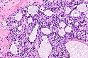
 Microcystic stromal tumour of ovary. H&E stain. | |
|
| |
| LM | microscopic cysts alternating with solid cellular regions, hyalinized fibrous stroma |
| LM DDx | other sex cord stromal tumours |
| IHC | CD10 +ve (cytoplasmic), beta-catenin (nuclear) +ve, WT-1 +ve, cyclin D1 +ve, inhibin -ve, calretinin -ve, ER-, PR- |
| Site | ovary, testis |
|
| |
| Prevalence | rare, evolving entity |
| Prognosis | unknown |
| Clin. DDx | other ovarian tumours, other testicular tumours |
| Treatment | excision |
Ovarian microcystic stromal tumour (abbreviated MST), also microcystic stromal tumour of the ovary, is a rare ovarian tumour.
General
- Evolving entity.
- Reported in the testis.[1]
- Sex cord stromal tumour.[2][3]
Microscopic
Features:[4]
- Microcysts alternating with solid cellular regions.
- Hyalinized fibrous stroma.
DDx:
- Other sex cord stromal tumours.
Images
IHC
Features:[4]
- CD10 +ve (cytoplasmic).
- Beta-catenin +ve (nuclear).
- WT1 +ve.
- Cyclin D1 +ve.
- Inhibin -ve.
- Calretinin -ve.
- ER -ve.
- PR -ve.
Molecular
- CTNNB1 mutation.[5]
References
- ↑ Zhu, P.; Duan, Y.; Ao, Q.; Wang, G. (Oct 2018). "Microcystic Stromal Tumor of Testicle: First Case Report and Literature Review.". Cancer Res Treat 50 (4): 1452-1457. doi:10.4143/crt.2017.414. PMID 29169232.
- ↑ Irving, JA.; Lee, CH.; Yip, S.; Oliva, E.; McCluggage, WG.; Young, RH. (Oct 2015). "Microcystic Stromal Tumor: A Distinctive Ovarian Sex Cord-Stromal Neoplasm Characterized by FOXL2, SF-1, WT-1, Cyclin D1, and β-catenin Nuclear Expression and CTNNB1 Mutations.". Am J Surg Pathol 39 (10): 1420-6. doi:10.1097/PAS.0000000000000482. PMID 26200099.
- ↑ Jeong, D.; Hakam, A.; Abuel-Haija, M.; Chon, HS. (Aug 2018). "Ovarian microcystic stromal tumor: Radiologic-pathologic correlation.". Gynecol Oncol Rep 25: 11-14. doi:10.1016/j.gore.2018.05.004. PMID 30014019.
- ↑ 4.0 4.1 McCluggage, WG.; Chong, AS.; Attygalle, AD.; Clarke, BA.; Chapman, W.; Rivera, B.; Foulkes, WD. (Feb 2019). "Expanding the morphological spectrum of ovarian microcystic stromal tumour.". Histopathology 74 (3): 443-451. doi:10.1111/his.13755. PMID 30325056.
- ↑ Na, K.; Kim, EK.; Jang, W.; Kim, HS. (Jun 2017). "CTNNB1 Mutations in Ovarian Microcystic Stromal Tumors: Identification of a Novel Deletion Mutation and the Use of Pyrosequencing to Identify Reported Point Mutation.". Anticancer Res 37 (6): 3249-3258. doi:10.21873/anticanres.11688. PMID 28551672.


