Difference between revisions of "Trichilemmoma"
Jump to navigation
Jump to search
(redirect) |
m |
||
| (4 intermediate revisions by one other user not shown) | |||
| Line 1: | Line 1: | ||
{{ Infobox diagnosis | |||
| Name = {{PAGENAME}} | |||
| Image = SkinTumors-P6190341.JPG | |||
| Width = | |||
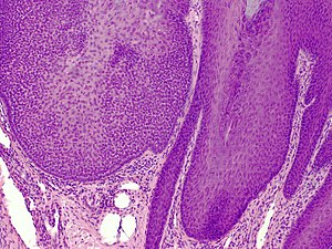
| Caption = Trichilemmoma. [[H&E stain]]. | |||
| Synonyms = | |||
| Micro = superficial dermal lesion contiguous with the epidermis; core of lesion has cuboidal cells with round nuclei, eosinophilic-clear cytoplasm; periphery of lesion surrounded by hyaline band, has peripheral palisading | |||
| Subtypes = | |||
| LMDDx = [[trichilemmal carcinoma]], [[basal cell carcinoma]], [[inverted follicular keratosis]] | |||
| Stains = | |||
| IHC = | |||
| EM = | |||
| Molecular = | |||
| IF = | |||
| Gross = | |||
| Grossing = | |||
| Site = [[skin]] | |||
| Assdx = | |||
| Syndromes = [[Cowden syndrome]] | |||
| Clinicalhx = | |||
| Signs = | |||
| Symptoms = | |||
| Prevalence = uncommon | |||
| Bloodwork = | |||
| Rads = | |||
| Endoscopy = | |||
| Prognosis = benign | |||
| Other = | |||
| ClinDDx = | |||
| Tx = | |||
}} | |||
'''Trichilemmoma''' is an uncommon [[Dermatologic neoplasms|skin tumour]]. | |||
==General== | |||
*Benign neoplasm with features of the pilosebaceous follicular epithelium.<ref>URL: [http://emedicine.medscape.com/article/1059940-overview http://emedicine.medscape.com/article/1059940-overview]. Accessed on: 2 September 2011.</ref> | |||
*Associated with ''[[nevus sebaceous]]''.<ref name=pmid16503928>{{Cite journal | last1 = Baykal | first1 = C. | last2 = Buyukbabani | first2 = N. | last3 = Yazganoglu | first3 = KD. | last4 = Saglik | first4 = E. | title = [Tumors associated with nevus sebaceous]. | journal = J Dtsch Dermatol Ges | volume = 4 | issue = 1 | pages = 28-31 | month = Jan | year = 2006 | doi = 10.1111/j.1610-0387.2006.05855.x | PMID = 16503928 }}</ref> | |||
*Muliple trichilemmomas associated with [[Cowden syndrome]].<ref name=Ref_Derm386>{{Ref Derm|386}}</ref> | |||
==Microscopic== | |||
Features:<ref name=Ref_Derm386>{{Ref Derm|386}}</ref> | |||
*Superficial dermal lesion contiguous with the epidermis: | |||
**Core of lesion: | |||
***Cuboidal cells with round nuclei, eosinophilic-clear cytoplasm. | |||
**Periphery of lesion: | |||
***Surrounded by hyaline band. | |||
***Peripheral palisading. | |||
DDx: | |||
*[[Trichilemmal carcinoma]]. | |||
*[[Basal cell carcinoma]]. | |||
*[[Inverted follicular keratosis]]. | |||
===Images=== | |||
*[http://dermimages.med.jhmi.edu/images/trichilemmoma_1_060109.jpg Trichilemmoma (jhmi.edu)].<ref>URL: [http://dermatlas.med.jhmi.edu/derm/indexDisplay.cfm?ImageID=667496720 http://dermatlas.med.jhmi.edu/derm/indexDisplay.cfm?ImageID=667496720]. Accessed on: 2 September 2011.</ref> | |||
*[http://www.flickr.com/photos/40981620@N04/3812019838/in/pool-1185084@N23/ Trichilemmoma - low mag. (flickr.com/Irlam)]. | |||
*[http://www.flickr.com/photos/40981620@N04/3812019930/in/pool-1185084@N23/ Trichilemmoma - intermed. mag. (flickr.com/Irlam)]. | |||
*[http://www.flickr.com/photos/40981620@N04/3811204517/in/pool-1185084@N23/ Trichilemmoma - high mag. (flickr.com/Irlam)]. | |||
==See also== | |||
*[[Dermatologic neoplasms]]. | |||
*[[Cowden syndrome]]. | |||
==References== | |||
{{Reflist|2}} | |||
[[Category:Diagnosis]] | |||
[[Category:Dermatologic neoplasms]] | |||
Latest revision as of 03:37, 20 February 2019
| Trichilemmoma | |
|---|---|
| Diagnosis in short | |
 Trichilemmoma. H&E stain. | |
|
| |
| LM | superficial dermal lesion contiguous with the epidermis; core of lesion has cuboidal cells with round nuclei, eosinophilic-clear cytoplasm; periphery of lesion surrounded by hyaline band, has peripheral palisading |
| LM DDx | trichilemmal carcinoma, basal cell carcinoma, inverted follicular keratosis |
| Site | skin |
|
| |
| Syndromes | Cowden syndrome |
|
| |
| Prevalence | uncommon |
| Prognosis | benign |
Trichilemmoma is an uncommon skin tumour.
General
- Benign neoplasm with features of the pilosebaceous follicular epithelium.[1]
- Associated with nevus sebaceous.[2]
- Muliple trichilemmomas associated with Cowden syndrome.[3]
Microscopic
Features:[3]
- Superficial dermal lesion contiguous with the epidermis:
- Core of lesion:
- Cuboidal cells with round nuclei, eosinophilic-clear cytoplasm.
- Periphery of lesion:
- Surrounded by hyaline band.
- Peripheral palisading.
- Core of lesion:
DDx:
Images
- Trichilemmoma (jhmi.edu).[4]
- Trichilemmoma - low mag. (flickr.com/Irlam).
- Trichilemmoma - intermed. mag. (flickr.com/Irlam).
- Trichilemmoma - high mag. (flickr.com/Irlam).
See also
References
- ↑ URL: http://emedicine.medscape.com/article/1059940-overview. Accessed on: 2 September 2011.
- ↑ Baykal, C.; Buyukbabani, N.; Yazganoglu, KD.; Saglik, E. (Jan 2006). "[Tumors associated with nevus sebaceous].". J Dtsch Dermatol Ges 4 (1): 28-31. doi:10.1111/j.1610-0387.2006.05855.x. PMID 16503928.
- ↑ 3.0 3.1 Busam, Klaus J. (2009). Dermatopathology: A Volume in the Foundations in Diagnostic Pathology Series (1st ed.). Saunders. pp. 386. ISBN 978-0443066542.
- ↑ URL: http://dermatlas.med.jhmi.edu/derm/indexDisplay.cfm?ImageID=667496720. Accessed on: 2 September 2011.