Difference between revisions of "Mantle cell lymphoma"
Jump to navigation
Jump to search
(→Images: added a case) |
|||
| (4 intermediate revisions by one other user not shown) | |||
| Line 43: | Line 43: | ||
==Microscopic== | ==Microscopic== | ||
Features:<ref>Good, D. 17 August 2010.</ref> | Features:<ref>Good, D. 17 August 2010.</ref> | ||
* | *Typically a small monomorphic (uniform size, shape and staining) lymphoid population. | ||
**May be "large" (blastic variant of mantle cell lymphoma) with 1 or 2 nucleoli.<ref name=pmid17917091>{{cite journal |author=Todorovic M, Pavlovic M, Balint B, ''et al.'' |title=Immunophenotypic profile and clinical characteristics in patients with advanced stage mantle cell lymphoma |journal=Med. Oncol. |volume=24 |issue=4 |pages=413–8 |year=2007 |pmid=17917091 |doi= |url=}}</ref> | |||
*Abundant mitoses. | *Abundant mitoses. | ||
*Scattered epithelioid histiocytes (should not be confused with tingible-body macrophages). | *Scattered epithelioid histiocytes (should not be confused with tingible-body macrophages). | ||
| Line 51: | Line 52: | ||
*Other [[small cell lymphomas]], esp. [[marginal zone lymphoma]]. | *Other [[small cell lymphomas]], esp. [[marginal zone lymphoma]]. | ||
*[[Burkitt's lymphoma]]. | *[[Burkitt's lymphoma]]. | ||
*Large B-cell lymphoma - for blastic variant of MCL.<ref name=pmid11681422>{{cite journal |author=Bernard M, Gressin R, Lefrère F, ''et al.'' |title=Blastic variant of mantle cell lymphoma: a rare but highly aggressive subtype |journal=Leukemia |volume=15 |issue=11 |pages=1785–91 |year=2001 |month=November |pmid=11681422 |doi= |url=}}</ref> | |||
===Images=== | ===Images=== | ||
| Line 61: | Line 63: | ||
www: | www: | ||
*[http://path.upmc.edu/cases/case441.html MCL - several images (upmc.edu)]. | *[http://path.upmc.edu/cases/case441.html MCL - several images (upmc.edu)]. | ||
[[File:4 718053471 sl 1.png|Mantle cell lymphoma]] | |||
[[File:4 718053471 sl 2.png|Mantle cell lymphoma]] | |||
[[File:4 718053471 sl 3.png|Mantle cell lymphoma]] | |||
[[File:4 718053471 sl 4.png|Mantle cell lymphoma]] | |||
[[File:4 718053471 sl 5.png|Mantle cell lymphoma]] | |||
[[File:4 718053471 sl 6.png|Mantle cell lymphoma]] | |||
[[File:4 718053471 sl 7.png|Mantle cell lymphoma]]<br> | |||
Mantle cell lymphoma in a 56 year old woman. A. A blue sea of cells spares a lymphoid follicle in the upper right corner. B. Small, round to reniform cells contrast with the larger tingible body macrophage (arrow). C-F. Tumor cells are Cyclin D1. CD20, BCL2, and CD5 positive positive. G. About 60% of tumor cells were positive for Ki67. Tumor cells were negative for CD3, CD10, and BCL6. | |||
==IHC== | ==IHC== | ||
| Line 78: | Line 88: | ||
==Molecular== | ==Molecular== | ||
*t(11;14)(q13;q32) / IGH-CCND1.<ref>URL: [http://atlasgeneticsoncology.org/Anomalies/t1114ID2021.html http://atlasgeneticsoncology.org/Anomalies/t1114ID2021.html]. Accessed on: 10 August 2010.</ref> | *t(11;14)(q13;q32) / IGH-CCND1.<ref>URL: [http://atlasgeneticsoncology.org/Anomalies/t1114ID2021.html http://atlasgeneticsoncology.org/Anomalies/t1114ID2021.html]. Accessed on: 10 August 2010.</ref> | ||
==Sign out== | |||
<pre> | |||
TERMINAL ILEUM, BIOPSY: | |||
- MANTLE CELL LYMPHOMA. | |||
COMMENT: | |||
The tumour consists of small lymphoid cells in sheets. | |||
The tumour stains as follows: | |||
POSITIVE: CD20, CD5, BCL2, cyclin D1. | |||
NEGATIVE: CD23, BCL6, CD10. | |||
PROLIFERATION (Ki-67): 25%. | |||
</pre> | |||
==See also== | ==See also== | ||
*[[Small cell lymphomas]]. | *[[Small cell lymphomas]]. | ||
*[[Ileal nodular lymphoid hyperplasia]]. | |||
==References== | ==References== | ||
Latest revision as of 20:28, 24 February 2017
| Mantle cell lymphoma | |
|---|---|
| Diagnosis in short | |
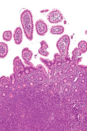
 Mantle cell lymphoma. H&E stain. | |
|
| |
| LM | small monomorphic (uniform size, shape and staining) lymphoid population with abundant mitoses, +/-scattered epithelioid histiocytes (should not be confused with tingible-body macrophages), +/-sclerosed blood vessels |
| Subtypes | blastic variant |
| LM DDx | other small cell lymphomas (esp. MALT lymphoma), Burkitt lymphoma |
| IHC | cyclin D1 +ve, CD5 +ve, CD43 +ve, CD20 +ve, CD23 -ve |
| Molecular | t(11;14)(q13;q32) / IGH-CCND1 |
| Site | lymph node, GI tract, other sites |
|
| |
| Prevalence | not common |
| Prognosis | moderately aggressive to poor |
Mantle cell lymphoma, abbreviated MCL, is less common small cell lymphoma that is relatively aggressive when compared to other small cell lymphomas.
General
- Relatively aggressive - guarded prognosis.[1]
- Rare ~ 2% of non-Hogkin's lymphoma in a series of over 4000 patients.[2]
GI tract - typically:[3]
- Abdominal pain (~37% of cases) or GI bleeding (~26% of cases).
Gross
- GI tract: polypoid lesions (~50% of cases).[3]
Microscopic
Features:[4]
- Typically a small monomorphic (uniform size, shape and staining) lymphoid population.
- May be "large" (blastic variant of mantle cell lymphoma) with 1 or 2 nucleoli.[5]
- Abundant mitoses.
- Scattered epithelioid histiocytes (should not be confused with tingible-body macrophages).
- Sclerosed blood vessels.
DDx:
- Other small cell lymphomas, esp. marginal zone lymphoma.
- Burkitt's lymphoma.
- Large B-cell lymphoma - for blastic variant of MCL.[6]
Images
www:


Mantle cell lymphoma in a 56 year old woman. A. A blue sea of cells spares a lymphoid follicle in the upper right corner. B. Small, round to reniform cells contrast with the larger tingible body macrophage (arrow). C-F. Tumor cells are Cyclin D1. CD20, BCL2, and CD5 positive positive. G. About 60% of tumor cells were positive for Ki67. Tumor cells were negative for CD3, CD10, and BCL6.
IHC
- CD45 +ve.
- CD20 +ve.
- CD79a +ve.
- CD5 +ve -- important.
- Negative in case reports.[7]
- CD43 +ve.
- Cyclin D1 +ve -- key immunostain.
Others:
- CD23 -ve.
- Positive in CLL.
Molecular
- t(11;14)(q13;q32) / IGH-CCND1.[9]
Sign out
TERMINAL ILEUM, BIOPSY:
- MANTLE CELL LYMPHOMA.

COMMENT:
The tumour consists of small lymphoid cells in sheets.

The tumour stains as follows:
POSITIVE: CD20, CD5, BCL2, cyclin D1.
NEGATIVE: CD23, BCL6, CD10.
PROLIFERATION (Ki-67): 25%.
See also
References
- ↑ Hankin, RC.; Hunter, SV. (Dec 1999). "Mantle cell lymphoma.". Arch Pathol Lab Med 123 (12): 1182-8. doi:10.1043/0003-9985(1999)1231182:MCL2.0.CO;2. PMID 10583923.
- ↑ Gujral S, Agarwal A, Gota V, et al. (2008). "A clinicopathologic study of mantle cell lymphoma in a single center study in India". Indian J Pathol Microbiol 51 (3): 315–22. PMID 18723950.
- ↑ 3.0 3.1 Kim, JH.; Jung, HW.; Kang, KJ.; Min, BH.; Lee, JH.; Chang, DK.; Kim, YH.; Son, HJ. et al. (2012). "Endoscopic findings in mantle cell lymphoma with gastrointestinal tract involvement.". Acta Haematol 127 (3): 129-34. doi:10.1159/000333139. PMID 22236942.
- ↑ Good, D. 17 August 2010.
- ↑ Todorovic M, Pavlovic M, Balint B, et al. (2007). "Immunophenotypic profile and clinical characteristics in patients with advanced stage mantle cell lymphoma". Med. Oncol. 24 (4): 413–8. PMID 17917091.
- ↑ Bernard M, Gressin R, Lefrère F, et al. (November 2001). "Blastic variant of mantle cell lymphoma: a rare but highly aggressive subtype". Leukemia 15 (11): 1785–91. PMID 11681422.
- ↑ URL: http://path.upmc.edu/cases/case308/dx.html. Accessed on: 14 January 2012.
- ↑ Online 'Mendelian Inheritance in Man' (OMIM) 168461
- ↑ URL: http://atlasgeneticsoncology.org/Anomalies/t1114ID2021.html. Accessed on: 10 August 2010.


