Difference between revisions of "Cholesteatoma"
Jump to navigation
Jump to search
| Line 54: | Line 54: | ||
- KERATINACEOUS DEBRIS AND GIANT CELLS, COMPATIBLE WITH CHOLESTEATOMA. | - KERATINACEOUS DEBRIS AND GIANT CELLS, COMPATIBLE WITH CHOLESTEATOMA. | ||
</pre> | </pre> | ||
===Micro=== | |||
The section shows keratinaceous debris and benign squamous epithelium with a granular layer. There is no significant cellular atypia and giant cells are not present. | |||
==See also== | ==See also== | ||
Revision as of 13:39, 29 December 2016
Cholesteatoma is a head and neck pathology ditzel.
General
- Squamous epithelium in the middle ear - leading to accumulation of keratinaceous debris.[1]
- The etiology is not well understood.[4][5]
- Theories include migration/hyperplasia, and metaplasia.[5]
- Rarely transforms into squamous cell carcinoma.[6][7]
Classification
May be subdivided into:[4]
- Acquired - due to trauma, surgery or infection.
- Congenital.
Gross
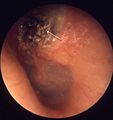
- Whitish mass in the middle ear.[8]
Image:
Microscopic
Features:[9]
- Keratinaceous debris - key feature.
- Squamous epithelium.
- Macrophages +/- giant cell (containing keratinceous debris).
- Chronic inflammation (lymphocytes).
DDx:
- Cholesterol granuloma.[10]
- Squamous cell carcinoma.[6]
- Epidermal inclusion cyst - history different.[citation needed]
Sign out
Submitted as "Left Mastoid Cholesteatoma", Excision:
	- Keratinaceous debris and benign squamous epithelium, consistent with cholesteatoma.
Soft tissue, left ear ("left ear keratosis"), excision:
	- Keratinaceous debris, squamous epithelium and bone (consistent with cholesteatoma).
Block letters
SOFT TISSUE (CHOLESTEATOMA), SITE NOT FURTHER SPECIFIED, REMOVAL:
	- KERATINACEOUS DEBRIS, COMPATIBLE WITH CHOLESTEATOMA.
TISSUE ("CHOLESTEATOMA"), LEFT, REMOVAL:
	- KERATINACEOUS DEBRIS AND GIANT CELLS, COMPATIBLE WITH CHOLESTEATOMA.
Micro
The section shows keratinaceous debris and benign squamous epithelium with a granular layer. There is no significant cellular atypia and giant cells are not present.
See also
References
- ↑ URL: http://www.harrisonspractice.com/practice/ub/view/Harrisons%20Practice/141015/all/otitis_media_and_mastoiditis. Accessed on: 16 March 2011.
- ↑ Piepergerdes MC, Kramer BM, Behnke EE (March 1980). "Keratosis obturans and external auditory canal cholesteatoma". Laryngoscope 90 (3): 383–91. PMID 7359960.
- ↑ Shire JR, Donegan JO (September 1986). "Cholesteatoma of the external auditory canal and keratosis obturans". Am J Otol 7 (5): 361–4. PMID 3538893.
- ↑ 4.0 4.1 Nevoux, J.; Lenoir, M.; Roger, G.; Denoyelle, F.; Ducou Le Pointe, H.; Garabédian, EN. (Sep 2010). "Childhood cholesteatoma.". Eur Ann Otorhinolaryngol Head Neck Dis 127 (4): 143-50. doi:10.1016/j.anorl.2010.07.001. PMID 20860924.
- ↑ 5.0 5.1 Louw, L. (Jun 2010). "Acquired cholesteatoma pathogenesis: stepwise explanations.". J Laryngol Otol 124 (6): 587-93. doi:10.1017/S0022215109992763. PMID 20156369.
- ↑ 6.0 6.1 Rothschild, S.; Ciernik, IF.; Hartmann, M.; Schuknecht, B.; Lütolf, UM.; Huber, AM.. "Cholesteatoma triggering squamous cell carcinoma: case report and literature review of a rare tumor.". Am J Otolaryngol 30 (4): 256-60. doi:10.1016/j.amjoto.2008.06.011. PMID 19563937.
- ↑ Takahashi, K.; Yamamoto, Y.; Sato, K.; Sato, Y.; Takahashi, S. (Jan 2005). "Middle ear carcinoma originating from a primary acquired cholesteatoma: a case report.". Otol Neurotol 26 (1): 105-8. PMID 15699729.
- ↑ Al Balushi, T.; Naik, JZ.; Al Khabori, M. (Jan 2013). "Congenital cholesteatoma in identical twins.". J Laryngol Otol 127 (1): 67-9. doi:10.1017/S0022215112002757. PMID 23217274.
- ↑ Iino Y, Toriyama M, Ohmi S, Kanegasaki S (1990). "Activation of peritoneal macrophages with human cholesteatoma debris and alpha-keratin". Acta Otolaryngol. 109 (5-6): 444–9. PMID 1694387.
- ↑ URL: http://path.upmc.edu/cases/case273/dx.html. Accessed on: 14 January 2012.
