Difference between revisions of "Primary sclerosing cholangitis"
(create) |
(→Images: Added a case) |
||
| (9 intermediate revisions by one other user not shown) | |||
| Line 1: | Line 1: | ||
'''Primary sclerosing cholangitis''', abbreviated '''PSC''', is an uncommon [[medical liver disease]] that can afflicts the young and old, and is often associated with [[ulcerative colitis]]. | |||
'''Pericholangitis''' is considered a synonym for ''small duct PSC''.<ref>URL: [http://emedicine.medscape.com/article/181889-overview http://emedicine.medscape.com/article/181889-overview]. Accessed on: 25 January 2012.</ref> | |||
==General== | |||
*Strongly associated with [[ulcerative colitis]]; 75-90% of PSC patients have [[inflammatory bowel disease]] (IBD).<ref name=emed_psc_over>Khurana V, Singh T. Primary sclerosing cholangitis. eMedicine.com. URL: [http://emedicine.medscape.com/article/187724-overview http://emedicine.medscape.com/article/187724-overview]. Accessed on: 29 November 2009.</ref> | |||
*Risk for [[cholangiocarcinoma]].<ref name=pmid19668124>{{Cite journal | last1 = Jesudian | first1 = AB. | last2 = Jacobson | first2 = IM. | title = Screening and diagnosis of cholangiocarcinoma in patients with primary sclerosing cholangitis. | journal = Rev Gastroenterol Disord | volume = 9 | issue = 2 | pages = E41-7 | month = | year = 2009 | doi = | PMID = 19668124 }}</ref> | |||
Serology: | |||
*p-ANCA (MPO-ANCA) +ve in ~ 90% of cases.<ref name=pmid11492972>{{Cite journal | last1 = Terjung | first1 = B. | last2 = Worman | first2 = HJ. | title = Anti-neutrophil antibodies in primary sclerosing cholangitis. | journal = Best Pract Res Clin Gastroenterol | volume = 15 | issue = 4 | pages = 629-42 | month = Aug | year = 2001 | doi = 10.1053/bega.2001.0209 | PMID = 11492972 }}</ref> | |||
===Diagnosis=== | |||
*'''Diagnosed radiologically'''. | |||
**Classically described as a ''chain of lakes''. | |||
*Liver biopsy is rarely useful diagnostically<ref name=emed_psc>Khurana V, Singh T. Primary sclerosing cholangitis. eMedicine.com. URL: [http://emedicine.medscape.com/article/187724-diagnosis http://emedicine.medscape.com/article/187724-diagnosis]. Accessed on: 29 November 2009.</ref> - as the disease may be patchy. | |||
**The utility of the biopsy is staging. | |||
===Treatment=== | |||
*None very good. | |||
*May be indication for transplant. | |||
==Microscopic== | |||
Features: | |||
*Classic: "onion-skinning" - cells layer around the bile ducts; "onion skin" present in approx. 40% of cases.<ref name=medscape_psc>Steele et al. URL: [http://www.medscape.com/viewarticle/552500_6 http://www.medscape.com/viewarticle/552500_6]. Accessed on: 29 November 2009.</ref> | |||
**Not pathognomonic for PSC<ref name=medscape_psc/> - but not too much else looks like this on microscopy (ergo good fellowship exam question). | |||
*+/-Ductopenia. | |||
*+/-Ductal proliferation. | |||
Notes: | |||
*PSC often has minimal inflammation.<ref>STC. 9 December 2010.</ref> | |||
DDx: | |||
*IgG4-associated cholangitis - see [[IgG4-related disease]].<ref name=pmid19633647>{{Cite journal | last1 = Deshpande | first1 = V. | last2 = Sainani | first2 = NI. | last3 = Chung | first3 = RT. | last4 = Pratt | first4 = DS. | last5 = Mentha | first5 = G. | last6 = Rubbia-Brandt | first6 = L. | last7 = Lauwers | first7 = GY. | title = IgG4-associated cholangitis: a comparative histological and immunophenotypic study with primary sclerosing cholangitis on liver biopsy material. | journal = Mod Pathol | volume = 22 | issue = 10 | pages = 1287-95 | month = Oct | year = 2009 | doi = 10.1038/modpathol.2009.94 | PMID = 19633647 }}</ref> | |||
*Others. | |||
===Staging=== | |||
Features:<ref>Steele et al. URL: [http://www.medscape.com/viewarticle/552500_6 http://www.medscape.com/viewarticle/552500_6]. Accessed on: 29 November 2009.</ref> | |||
*Stage I - focal portal inflammation, +/- duct abnormalities, no fibrosis. | |||
*Stage II - portal enlargement (fibrosis), +/- inflammation. | |||
*Stage III - bridging fibrosis + [[necrosis]]. | |||
*Stage IV - cirrhosis. | |||
Notes: | |||
*Similar to [[PBC]] staging. | |||
===Images=== | |||
*[http://trialx.com/curetalk/wp-content/blogs.dir/7/files/2011/05/diseases/Primary_Sclerosing_Cholangitis-2.jpg PSC (trialx.com)].<ref>URL: [http://trialx.com/curebyte/2011/07/08/clinical-trials-and-images-of-primary-sclerosing-cholangitis/ http://trialx.com/curebyte/2011/07/08/clinical-trials-and-images-of-primary-sclerosing-cholangitis/]. Accessed on: 1 January 2012.</ref> | |||
*[http://www.onmedica.com/getresource.aspx?resourceid=e398a3f2-e030-40e6-823b-2e8e2cbbe4bb PSC (onmedica.com)].<ref>URL: [http://www.onmedica.com/NewsArticle.aspx?id=d7f992b5-6dee-46c6-8383-bbdfb4528ccc http://www.onmedica.com/NewsArticle.aspx?id=d7f992b5-6dee-46c6-8383-bbdfb4528ccc]. Accessed on: 1 January 2011.</ref> | |||
*[http://www.nature.com/nrgastro/journal/v7/n11/fig_tab/nrgastro.2010.155_F2.html PSC (nature.com)].<ref>{{Cite journal | last1 = Mendes | first1 = F. | last2 = Lindor | first2 = KD. | title = Primary sclerosing cholangitis: overview and update. | journal = Nat Rev Gastroenterol Hepatol | volume = 7 | issue = 11 | pages = 611-9 | month = Nov | year = 2010 | doi = 10.1038/nrgastro.2010.155 | PMID = 20938459 }}</ref> | |||
{| | |||
[[File:1 PSC 1 680x512px.tif| Primary sclerosing cholangitis in patient with history of ulcerative colitis.]] | |||
[[File:2 PSC 1 680x512px.tif| Primary sclerosing cholangitis in patient with history of ulcerative colitis.]] | |||
<br> | |||
[[File:3 PSC 1 680x512px.tif| Primary sclerosing cholangitis in patient with history of ulcerative colitis.]] | |||
[[File:4 PSC 1 680x512px.tif| Primary sclerosing cholangitis in patient with history of ulcerative colitis.]] | |||
<br> | |||
[[File:5 PSC 1 680x512px.tif| Primary sclerosing cholangitis in patient with history of ulcerative colitis.]] | |||
[[File:6 PSC 1 680x512px.tif| Primary sclerosing cholangitis in patient with history of ulcerative colitis.]] | |||
|} | |||
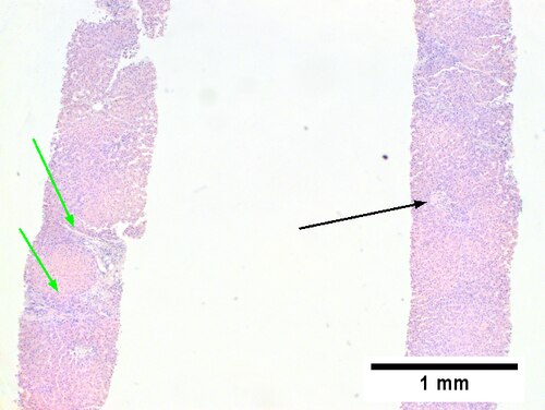
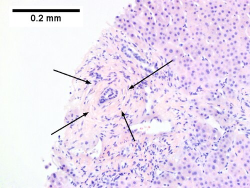
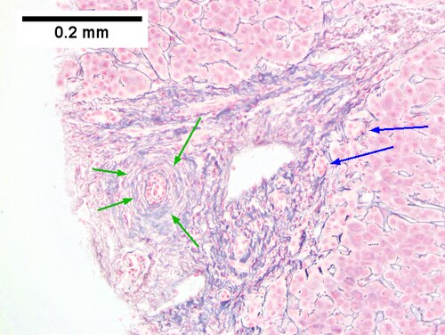
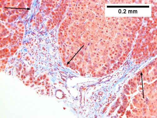
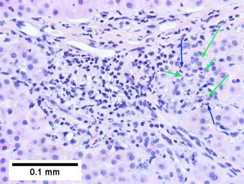
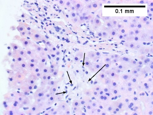
<br>Primary sclerosing cholangitis in patient with history of ulcerative colitis. <br>Low power shows inflamed bands [green arrows] and a rare scar at the edge of a triad [black arrow] (Row 1 Left 40X). A triad with a characteristic periductal fibrosis [black arrows] is seen (Row 1 Right 200X). Reticulin shows the space about the duct [green arrows], as well as some foci of piecemeal necrosis [blue arrows] (Row 2 Left 200X). Trichrome shows fibrous bridges [black arrows] (Row 2 Right 200X). Damage to bile ducts is seen by peripheral proliferated bile ductules [green arrows] with neutrophils [blue arrows] (Row 3 Left 400X). Also seen is a small periportal bile infarct with necrotic hepatocyte nuclei [black arrows] (Row 3 Right 400X). | |||
==See also== | |||
*[[Medical liver disease]]. | |||
==References== | |||
{{Reflist|2}} | |||
[[Category:Diagnosis]] | |||
[[Category:Medical liver disease]] | |||
Latest revision as of 21:21, 11 September 2016
Primary sclerosing cholangitis, abbreviated PSC, is an uncommon medical liver disease that can afflicts the young and old, and is often associated with ulcerative colitis.
Pericholangitis is considered a synonym for small duct PSC.[1]
General
- Strongly associated with ulcerative colitis; 75-90% of PSC patients have inflammatory bowel disease (IBD).[2]
- Risk for cholangiocarcinoma.[3]
Serology:
- p-ANCA (MPO-ANCA) +ve in ~ 90% of cases.[4]
Diagnosis
- Diagnosed radiologically.
- Classically described as a chain of lakes.
- Liver biopsy is rarely useful diagnostically[5] - as the disease may be patchy.
- The utility of the biopsy is staging.
Treatment
- None very good.
- May be indication for transplant.
Microscopic
Features:
- Classic: "onion-skinning" - cells layer around the bile ducts; "onion skin" present in approx. 40% of cases.[6]
- Not pathognomonic for PSC[6] - but not too much else looks like this on microscopy (ergo good fellowship exam question).
- +/-Ductopenia.
- +/-Ductal proliferation.
Notes:
- PSC often has minimal inflammation.[7]
DDx:
- IgG4-associated cholangitis - see IgG4-related disease.[8]
- Others.
Staging
Features:[9]
- Stage I - focal portal inflammation, +/- duct abnormalities, no fibrosis.
- Stage II - portal enlargement (fibrosis), +/- inflammation.
- Stage III - bridging fibrosis + necrosis.
- Stage IV - cirrhosis.
Notes:
- Similar to PBC staging.
Images
Primary sclerosing cholangitis in patient with history of ulcerative colitis.
Low power shows inflamed bands [green arrows] and a rare scar at the edge of a triad [black arrow] (Row 1 Left 40X). A triad with a characteristic periductal fibrosis [black arrows] is seen (Row 1 Right 200X). Reticulin shows the space about the duct [green arrows], as well as some foci of piecemeal necrosis [blue arrows] (Row 2 Left 200X). Trichrome shows fibrous bridges [black arrows] (Row 2 Right 200X). Damage to bile ducts is seen by peripheral proliferated bile ductules [green arrows] with neutrophils [blue arrows] (Row 3 Left 400X). Also seen is a small periportal bile infarct with necrotic hepatocyte nuclei [black arrows] (Row 3 Right 400X).
See also
References
- ↑ URL: http://emedicine.medscape.com/article/181889-overview. Accessed on: 25 January 2012.
- ↑ Khurana V, Singh T. Primary sclerosing cholangitis. eMedicine.com. URL: http://emedicine.medscape.com/article/187724-overview. Accessed on: 29 November 2009.
- ↑ Jesudian, AB.; Jacobson, IM. (2009). "Screening and diagnosis of cholangiocarcinoma in patients with primary sclerosing cholangitis.". Rev Gastroenterol Disord 9 (2): E41-7. PMID 19668124.
- ↑ Terjung, B.; Worman, HJ. (Aug 2001). "Anti-neutrophil antibodies in primary sclerosing cholangitis.". Best Pract Res Clin Gastroenterol 15 (4): 629-42. doi:10.1053/bega.2001.0209. PMID 11492972.
- ↑ Khurana V, Singh T. Primary sclerosing cholangitis. eMedicine.com. URL: http://emedicine.medscape.com/article/187724-diagnosis. Accessed on: 29 November 2009.
- ↑ 6.0 6.1 Steele et al. URL: http://www.medscape.com/viewarticle/552500_6. Accessed on: 29 November 2009.
- ↑ STC. 9 December 2010.
- ↑ Deshpande, V.; Sainani, NI.; Chung, RT.; Pratt, DS.; Mentha, G.; Rubbia-Brandt, L.; Lauwers, GY. (Oct 2009). "IgG4-associated cholangitis: a comparative histological and immunophenotypic study with primary sclerosing cholangitis on liver biopsy material.". Mod Pathol 22 (10): 1287-95. doi:10.1038/modpathol.2009.94. PMID 19633647.
- ↑ Steele et al. URL: http://www.medscape.com/viewarticle/552500_6. Accessed on: 29 November 2009.
- ↑ URL: http://trialx.com/curebyte/2011/07/08/clinical-trials-and-images-of-primary-sclerosing-cholangitis/. Accessed on: 1 January 2012.
- ↑ URL: http://www.onmedica.com/NewsArticle.aspx?id=d7f992b5-6dee-46c6-8383-bbdfb4528ccc. Accessed on: 1 January 2011.
- ↑ Mendes, F.; Lindor, KD. (Nov 2010). "Primary sclerosing cholangitis: overview and update.". Nat Rev Gastroenterol Hepatol 7 (11): 611-9. doi:10.1038/nrgastro.2010.155. PMID 20938459.