|
|
| Line 1: |
Line 1: |
| '''Soft tissue lesions''' strike fear in many pathologists as they are uncommon and may be difficult to diagnose. | | {{ Infobox diagnosis |
| | | Name = {{PAGENAME}} |
| | | Image = Alveolar_soft_part_sarcoma_-2-_very_high_mag.jpg |
| | | Width = |
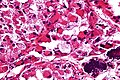
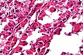
| | | Caption = ASPS. [[H&E stain]]. |
| | | Micro = large cells (~30-50 μm) with abundant eosinophilic cytoplasm and an eccentric nucleus +/-nucleolus, arranged in nests/separated by thin septa - vaguely resembles alveoli (at low power) |
| | | Subtypes = |
| | | LMDDx = [[paraganglioma]], [[clear cell renal cell carcinoma]], [[alveolar rhabdomyosarcoma]] |
| | | Stains = PAS +ve (cytoplasm) |
| | | IHC = TFE3 +ve (suggestive of translocation) |
| | | EM = intracytoplasmic crystal lattice with ~5 nm fibres spaced 10 nm apart |
| | | Molecular = t(X;17) |
| | | IF = |
| | | Gross = |
| | | Grossing = |
| | | Site = [[soft tissue lesions|soft tissue]] - usu. [[head and neck pathology|head and neck]] |
| | | Assdx = |
| | | Syndromes = |
| | | Clinicalhx = usu. slow growing mass |
| | | Signs = |
| | | Symptoms = |
| | | Prevalence = rare |
| | | Bloodwork = |
| | | Rads = |
| | | Endoscopy = |
| | | Prognosis = ultimately poor |
| | | Other = |
| | | ClinDDx = |
| | }} |
| | '''Alveolar soft part sarcoma''', abbreviated '''ASPS''', is a rare [[malignant]] soft tissue lesion typically seen in younger individuals. |
|
| |
|
| =Introduction=
| | ==General== |
| ==WHO classification of soft tissue lesions/tumours==
| |
| ===Morphologic grouping<ref name=Ref_WMSP601-3>{{Ref WMSP|601-3}}</ref>===
| |
| #Adipocytic tumours.
| |
| #Fibroblastic/myofibroblastic tumours.
| |
| #"Fibrohistiocytic" tumours.
| |
| #Smooth muscle tumours.
| |
| #Skeletal muscle tumours.
| |
| #Vascular tumours.
| |
| #Perivascular (pericytic) tumours.
| |
| #Chondro-osseous tumours.
| |
| #Tumours of uncertain differentiation.
| |
| | |
| ===Biologic potential grouping<ref>{{Ref WMSP|598-604}}</ref>===
| |
| #Benign.
| |
| #Intermediate (locally aggressive).
| |
| #Intermediate (rarely metastasizing).
| |
| #Malignant.
| |
| | |
| ==Prevalence==
| |
| *All sarcomas are rare buggers.
| |
| **As the classification has been changing over the past years (with more subtypes being recognized/identified) numbers are variable from study-to-study.
| |
| *Once upon a time almost everything was called ''malignant fibrous histiocytoma''; thus, it is listed as a common entity in some publications.
| |
| | |
| ===Most common:<ref name=pmid17976362>{{cite journal |author=Skubitz KM, D'Adamo DR |title=Sarcoma |journal=Mayo Clin. Proc. |volume=82 |issue=11 |pages=1409–32 |year=2007 |month=November |pmid=17976362 |doi= |url= http://www.mayoclinicproceedings.com/content/82/11/1409.long}}</ref>===
| |
| *Liposarcoma.
| |
| *Leiomyosarcoma.
| |
| | |
| ==Molecular testing==
| |
| {{Main|Molecular pathology}}
| |
| *Molecular testing plays an important role in soft tissue pathology.
| |
| *It is generally seen as an adjunct test that:<ref name=pmid11454050>{{cite journal |author=Fletcher CD, Fletcher JA, Dal Cin P, Ladanyi M, Woodruff JM |title=Diagnostic gold standard for soft tissue tumours: morphology or molecular genetics? |journal=Histopathology |volume=39 |issue=1 |pages=100–3 |year=2001 |month=July |pmid=11454050 |doi= |url=}}</ref>
| |
| **Often is used to confirm the histomorphologic impression/quality control.
| |
| **Frequently has some prognostic significance.
| |
| **May directly affect treatment.
| |
| | |
| ===Translocations===
| |
| {{Main|Chromosomal translocations}}
| |
| *Many tumours in soft tissue pathology are diagnosed inconjunction with the finding of [[chromosomal translocations]].
| |
| | |
| ==Histologic patterns==
| |
| {| class="wikitable sortable" style="margin-left:auto;margin-right:auto"
| |
| ! Name
| |
| ! Description
| |
| ! DDx
| |
| ! Image
| |
| ! Other
| |
| |-
| |
| | Storiform, [[AKA]] patternless pattern<ref name=pmid9704618>{{cite journal |author=Mangano WE, Cagle PT, Churg A, Vollmer RT, Roggli VL |title=The diagnosis of desmoplastic malignant mesothelioma and its distinction from fibrous pleurisy: a histologic and immunohistochemical analysis of 31 cases including p53 immunostaining |journal=Am. J. Clin. Pathol. |volume=110 |issue=2 |pages=191–9 |year=1998 |month=August |pmid=9704618 |doi= |url=}}</ref>
| |
| | whorled, cartwheel-like arrangement
| |
| | [[Undifferentiated pleomorphic sarcoma]]
| |
| | image ?
| |
| | other ?
| |
| |-
| |
| | Herring bone
| |
| | like herring bone (technique) for climbing a hill in cross country skiing; books on a shelf, where they have partially fallen over -- on the one shelf to the left and the one below to the right
| |
| | [[fibrosarcoma]], [[synovial sarcoma]], [[MPNST]]
| |
| | image ?
| |
| | other ?
| |
| |-
| |
| | Fasicular
| |
| | the long axis of the (spindle) cells are perpendicular to one another in adjacent bundles of cells
| |
| | [[leiomyoma]]
| |
| | image ?
| |
| | other ?
| |
| |-
| |
| | Biphasic
| |
| | nests of cells and stroma
| |
| | [[synovial sarcoma]], [[DSRCT]], [[alveolar RMS]]
| |
| | image ?
| |
| | other ?
| |
| |- <!--
| |
| | name ?
| |
| | description ?
| |
| | DDx ?
| |
| | image ?
| |
| | other ? -->
| |
| |}
| |
| | |
| =Adipocytic tumours=
| |
| {{Main|Adipocytic tumours}}
| |
| | |
| This category includes:
| |
| *Lipoma.
| |
| *Liposarcoma.
| |
| *Hibernoma.
| |
| | |
| =Smooth muscle tumours=
| |
| ==Leiomyosarcoma==
| |
| See gyne notes.
| |
| | |
| ===Microscopy===
| |
| Features:
| |
| *Nuclear atypia.
| |
| *[[Necrosis]].
| |
| *Mitoses.
| |
| | |
| =Fibrohistiocytic tumours=
| |
| ==Pleomorphic undifferentiated sarcoma==
| |
| *Abbreviated ''PUS''.
| |
| *[[AKA]] ''Undifferentiated pleomorphic sarcoma''.
| |
| *Previously known as ''malignant fibrous histiocytoma'', abbreviated ''MFH''.<ref>URL: [http://sarcomahelp.org/learning_center/mfh.html http://sarcomahelp.org/learning_center/mfh.html]. Accessed on: 8 April 2011.</ref>
| |
| | |
| ===General===
| |
| *Common sarcoma.
| |
| *Usu. deep tissue of the trunk and extremities.
| |
| | |
| ===Microscopic===
| |
| Features:<ref name=Ref_WMSP_613>{{Ref WMSP|613}}</ref>
| |
| *Storiform pattern ([[AKA]] ''patternless pattern'') - '''key feature'''.
| |
| *Marked nuclear pleomorphism '''key feature'''.
| |
| **Variation is nuclear size, nuclear shape and nuclear staining (esp. hyperchromasia).
| |
| *Mitoses - abundant; atypical mitoses common.
| |
| *Necrosis (common).
| |
| *Mix of spindle cells and epithelioid cells.
| |
| | |
| Other findings:
| |
| *+/-Giant cells (see subclassification).
| |
| *+/-Inflammation (see subclassification).
| |
| **Neutrophils.
| |
| **Eosinophils.
| |
| | |
| ====Subclassification====
| |
| Pleomorphic sarcoma (PS) is subclassified the following way:<ref name=Ref_WMSP_613-4>{{Ref WMSP|613-4}}</ref>
| |
| *PS with giant cells.
| |
| *PS with inflammation.
| |
| *PUS (not otherwise specified) - wastebasket diagnosis; if neither of the above two apply.
| |
| | |
| =Fibroblastic/myofibroblastic tumours=
| |
| | |
| ==Proliferative fasciitis==
| |
| *Need to write something here.
| |
| | |
| ==Solitary fibrous tumour==
| |
| ===General===
| |
| *Grouped with ''hemangiopericytoma'' in the WHO classification; possibly the same tumour (?).<ref name=Ref_WMSP609>{{Ref WMSP|609}}</ref>
| |
| *May be benign ''or'' malignant; more commonly benign.<ref>URL: [http://www.pathconsultddx.com/pathCon/diagnosis?pii=S1559-8675%2806%2970528-9 http://www.pathconsultddx.com/pathCon/diagnosis?pii=S1559-8675%2806%2970528-9]. Accessed on: 25 June 2010.</ref><ref>URL: [http://wjso.com/content/6/1/86 http://wjso.com/content/6/1/86]. Accessed on: 25 June 2010.</ref>
| |
| | |
| ===Microscopic===
| |
| Features:
| |
| *Well-circumscribed.
| |
| *Fibroblast-like cells (spindle cells).
| |
| *Hemangiopericytoma-like area (staghorn vessels) - not seen on image.
| |
| *Keloid-like collagen bundles.
| |
| | |
| Images:
| |
| *[http://commons.wikimedia.org/wiki/File:Solitary_fibrous_tumour_low_mag.jpg SFT - low mag. (WC)].
| |
| *[http://commons.wikimedia.org/wiki/File:Solitary_fibrous_tumour_intermed_mag.jpg SFT - intermed. mag. (WC)].
| |
| *[http://commons.wikimedia.org/wiki/File:Solitary_fibrous_tumour_high_mag.jpg SFT - high mag. (WC)].
| |
| | |
| ===IHC===
| |
| *CD34 ~90% +ve.
| |
| *CD99 ~70% +ve.
| |
| *BCL2 ~50% +ve.
| |
| | |
| ==Hemangiopericytoma==
| |
| ===General===
| |
| *Grouped with ''solitary fibrous tumour'' in the WHO classification; possibly the same tumour (?).<ref name=Ref_WMSP609>{{Ref WMSP|609}}</ref>
| |
| *Arises from the ''pericyte'', a connective tissue cell of small vessels that is thought to be involved in flow regulation.
| |
| *Hematologic spread most common - to lungs.<ref>URL: [http://emedicine.medscape.com/article/1255879-overview http://emedicine.medscape.com/article/1255879-overview]. Accessed on: 2 May 2010.</ref>
| |
| *Oncogenic osteomalacia - assoc. with hemangiopericytoma.<ref>URL: [http://emedicine.medscape.com/article/1255879-overview http://emedicine.medscape.com/article/1255879-overview]. Accessed on: 2 May 2010.</ref>
| |
| | |
| ===Presentation===
| |
| *Usually painless mass, slow enlargement.
| |
| | |
| ====Radiology====
| |
| *Intramedullary lytic mass.
| |
| *May be well-circumscribed.
| |
| *+/-Periosteal reaction.
| |
| *+/-Sclerotic border.
| |
| | |
| May be worked-up with angiography to distinguish from a [[vascular malformation]].<ref>URL: [http://emedicine.medscape.com/article/1255879-diagnosis http://emedicine.medscape.com/article/1255879-diagnosis]. Accessed on: 2 May 2010.</ref>
| |
| ===Location===
| |
| *Usually extremities - femur or prox. tibial.<ref>URL: [http://emedicine.medscape.com/article/1255879-overview http://emedicine.medscape.com/article/1255879-overview]. Accessed on: 2 May 2010.</ref>
| |
| | |
| ===Histology===
| |
| Features:<ref>URL: [http://emedicine.medscape.com/article/1255879-diagnosis http://emedicine.medscape.com/article/1255879-diagnosis]. Accessed on: 2 May 2010.</ref>
| |
| *Hypervascular lesion - '''key diagnostic feature'''.<ref name=enzinger>Enzinger & Weiss's Soft Tissue Tumors. 4th Ed. PP.1007-13. ISBN 0-323-01200-0.</ref>
| |
| **Abundant thin-walled branching small vessels of variable size.
| |
| ***May be described as "staghorn vessels" or "antler-like" vasculature.
| |
| ***Cells may "onion-skin" around thin blood vessels.
| |
| *Spindle or ovoid shaped cells in nests or sheets.
| |
| | |
| ===IHC===
| |
| Features:<ref name=Ref_WMSP609>{{Ref WMSP|609}}</ref><ref name=enzinger/>
| |
| *Vimentin +ve (usually).
| |
| *Desmin -ve (typical).
| |
| *Factor VIII -ve (marks endothelium).
| |
| *CD34 +ve.
| |
| **CD34 usu. -ve in synovial sarcoma.
| |
| *CD31 -ve (marks benign endothelium).
| |
| *vWF (von Willebrand factor) -ve.
| |
| | |
| May be in the DDx for [[meningioma]]:<ref>Croul, SE. 8 November 2010.</ref>
| |
| *EMA -ve.
| |
| *S100 -ve.
| |
| | |
| ===DDx===
| |
| *Other vascular tumours.
| |
| *Vascular malformations.
| |
| *Synovial sarcoma.
| |
| | |
| ==Desmoplastic fibroblastoma==
| |
| *AKA ''collagenous fibroma''.<ref name=pmid18271804>{{Cite journal | last1 = Watanabe | first1 = H. | last2 = Ishida | first2 = Y. | last3 = Nagashima | first3 = K. | last4 = Makino | first4 = T. | last5 = Norisugi | first5 = O. | last6 = Shimizu | first6 = T. | title = Desmoplastic fibroblastoma (collagenous fibroma). | journal = J Dermatol | volume = 35 | issue = 2 | pages = 93-7 | month = Feb | year = 2008 | doi = 10.1111/j.1346-8138.2008.00421.x | PMID = 18271804 }}</ref>
| |
| *Benign lesion.
| |
| *Classically found in shoulder region.
| |
| | |
| ===IHC===
| |
| *Beta-catenin -ve.<ref name=pmid18544056>{{Cite journal | last1 = Takahara | first1 = M. | last2 = Ichikawa | first2 = R. | last3 = Oda | first3 = Y. | last4 = Uchi | first4 = H. | last5 = Takeuchi | first5 = S. | last6 = Moroi | first6 = Y. | last7 = Kiryu | first7 = H. | last8 = Furue | first8 = M. | title = Desmoplastic fibroblastoma: a case presenting as a protruding nodule in the dermis. | journal = J Cutan Pathol | volume = 35 Suppl 1 | issue = | pages = 70-3 | month = Oct | year = 2008 | doi = 10.1111/j.1600-0560.2007.00964.x | PMID = 18544056 }}
| |
| </ref>
| |
| **Significance ???
| |
| | |
| ==Low-grade fibromyxoid sarcoma==
| |
| *[[AKA]] ''hyalinizing spindle cell tumour''.
| |
| ===General===
| |
| *Deep soft tissue.
| |
| | |
| ===Microscopic===
| |
| Features:<ref>{{cite journal |author=Vernon SE, Bejarano PA |title=Low-grade fibromyxoid sarcoma: a brief review |journal=Arch. Pathol. Lab. Med. |volume=130 |issue=9 |pages=1358–60 |year=2006 |month=September |pmid=16948525 |doi= |url=}}</ref>
| |
| *Myoid stroma - '''key feature'''.
| |
| *Low cellularity.
| |
| *Spindle cells.
| |
| | |
| Notes:
| |
| *Few/absent mitoses.
| |
| | |
| ===Molecular pathology===
| |
| t(7;16)(q33;p11)<ref>{{cite journal |author=Panagopoulos I, Storlazzi CT, Fletcher CD, ''et al.'' |title=The chimeric FUS/CREB3l2 gene is specific for low-grade fibromyxoid sarcoma |journal=Genes Chromosomes Cancer |volume=40 |issue=3 |pages=218–28 |year=2004 |month=July |pmid=15139001 |doi=10.1002/gcc.20037 |url=}}</ref>
| |
| | |
| =Vascular lesions=
| |
| Vascular lesions are "too red"; they have too many RBCs.
| |
| | |
| ==Hemangioma==
| |
| ===General===
| |
| Comes is various flavours:<ref name=pmid10410855>{{cite journal |author=Prieto VG, Shea CR |title=Selected cutaneous vascular neoplasms. A review |journal=Dermatol Clin |volume=17 |issue=3 |pages=507–20, viii |year=1999 |month=July |pmid=10410855 |doi= |url=}}</ref>
| |
| *Tufted.
| |
| **Small clusters of blood vessels.
| |
| *Microvenular hemangioma.
| |
| *Glomeruloid hemangioma - associated with [[POEMS syndrome]].
| |
| *Epithelioid hemangioma.
| |
| *Targetoid hemosideric hemangioma.
| |
| | |
| ===Microscopic===
| |
| Features:
| |
| *Abundance of benign small blood vessels. (???)
| |
| | |
| ==Kaposi sarcoma==
| |
| ===General===
| |
| *Not really a sarcoma.
| |
| *Caused by HHV-8.
| |
| *Associated with immunodeficiency, e.g. [[HIV]]/AIDS.
| |
| | |
| ===Stages===
| |
| It is seen in different stages:<ref>URL: [http://www.histopathology-india.net/KS.htm http://www.histopathology-india.net/KS.htm]. Accessed on: 31 January 2010.</ref>
| |
| #Patch stage.
| |
| #Plaque stage.
| |
| #Nodular stage.
| |
| #Lymphangioma-like. (???)
| |
| | |
| ===Microscopic===
| |
| Features:<ref name=Ref_Klatt23>{{Ref Klatt|23}}</ref>- '''key feature'''.
| |
| *+/-Nuclear atypia.
| |
| *Hyaline globules (intracytoplasmic)<ref name=pmid7528163>{{cite journal |author=del Rosario AD, Bui HX, Singh J, Ginsburg R, Ross JS |title=Intracytoplasmic eosinophilic hyaline globules in cartilaginous neoplasms: a surgical, pathological, ultrastructural, and electron probe x-ray microanalytic study |journal=Hum. Pathol. |volume=25 |issue=12 |pages=1283–9 |year=1994 |month=December |pmid=7528163 |doi= |url=}}</ref> - pale pink globs (that are paler than RBCs) - '''important feature'''.
| |
| *+/-Hemosiderin deposits.
| |
| | |
| DDx:
| |
| *Angiosarcoma (have many mitoses, nuclear atypia).
| |
| *Masson's hemangioma (Intravascular papillary endothelial hyperplasia).
| |
| | |
| Notes:
| |
| *Hyaline globules have a DDx ([[hepatocellular carcinoma]], lung adenocarcinoma, [[chondrosarcoma]]s + others).<ref name=pmid7528163/>
| |
| *Promontory sign - small vessel protruding into an abnormal vascular space.<ref name=pmid19318797>{{cite journal |author=Lazova R, McNiff JM, Glusac EJ, Godic A |title=Promontory sign--present in patch and plaque stage of angiosarcoma! |journal=Am J Dermatopathol |volume=31 |issue=2 |pages=132–6 |year=2009 |month=April |pmid=19318797 |doi=10.1097/DAD.0b013e3181951045 |url=}}</ref>
| |
| **Not pathognomonic for KS.<ref name=pmid20577080>{{cite journal |author=Fernandez-Flores A, Rodriguez R |title=Promontory Sign in a Reactive Benign Vascular Proliferation |journal=Am J Dermatopathol |volume= |issue= |pages= |year=2010 |month=June |pmid=20577080 |doi=10.1097/DAD.0b013e3181cf0ae5 |url=}}</ref>
| |
| **Image: [http://www.diagnosticpathology.org/content/3/1/31/figure/F1 Promontory sign (diagnosticpathology.org)].
| |
| | |
| Images:
| |
| *[http://commons.wikimedia.org/wiki/File:Kaposi_sarcoma_high_mag.jpg Kaposi sacoma - high mag. (WC)].
| |
| *[http://commons.wikimedia.org/wiki/File:Kaposi_sarcoma_low_intermed_mag.jpg Kaposi sarcoma - intermed. mag. (WC)].
| |
| | |
| ===IHC===
| |
| *CD31 +ve.
| |
| *CD34 +ve.
| |
| *HHV-8 +ve.
| |
| | |
| ==Masson hemangioma==
| |
| ===General===
| |
| *Benign non-neoplastic lesion - a vessel that has thrombosed and recanalized.
| |
| *[[AKA]] ''intravascular papillary endothelial hyperplasia''.<ref name=pmid15865098>{{cite journal |author=Korkolis DP, Papaevangelou M, Koulaxouzidis G, Zirganos N, Psichogiou H, Vassilopoulos PP |title=Intravascular papillary endothelial hyperplasia (Masson's hemangioma) presenting as a soft-tissue sarcoma |journal=Anticancer Res. |volume=25 |issue=2B |pages=1409–12 |year=2005 |pmid=15865098 |doi= |url=}}</ref>
| |
| *Histomorphologically may be confused with low-grade angiosarcoma or other soft tissue sarcomas.<ref name=pmid15865098/>
| |
| ===Microscopic===
| |
| Features:
| |
| *Well-circumscribed - '''key (low power) feature'''.
| |
| *Abundant small vascular channels with benign endothelium.
| |
| | |
| Notes:
| |
| *Looks like Kaposi sarcoma at high power.
| |
| | |
| ==Angiosarcoma==
| |
| ===General===
| |
| *Malignant tumour - with a horrible prognosis.<ref name=pmid20537949>{{cite journal |author=Young RJ, Brown NJ, Reed MW, Hughes D, Woll PJ |title=Angiosarcoma |journal=Lancet Oncol |volume= |issue= |pages= |year=2010 |month=May |pmid=20537949 |doi=10.1016/S1470-2045(10)70023-1 |url=}}</ref>
| |
| *Classically on the scalp or head & neck.
| |
| *May arise secondary to therapeutic radiation or chronic lymphoedema related to breast carcinoma.
| |
| | |
| ===Microscopic===
| |
| Features:
| |
| *Very many small capillaries of irregular shape lined with:
| |
| **Pleomorphic nuclei.
| |
| ***May have hobnail morphology.
| |
| *Mitoses.
| |
| *Cytoplasmic vacuoles.
| |
| **Cells trying to form lumina - embryologic.
| |
| | |
| Notes:
| |
| *Epithelioid variant (with abundant cytoplasm & sheeting architecture) may resemble ''[[melanoma]]'' or ''[[hepatocellular carcinoma]]''.
| |
| | |
| ===IHC===
| |
| *CD34 +ve.
| |
| *D2-40 +ve. (???)
| |
| *CD31 +ve.
| |
| | |
| ==Hemangioendothelioma==
| |
| ===General===
| |
| *Usually benign.
| |
| | |
| ===Microscopic===
| |
| Features:<ref name=Ref_Klatt23>{{Ref Klatt|23}}</ref>
| |
| *Well-formed thin vascular channels on a fibrous stroma - '''key feature'''.
| |
| *+/-Thrombosis.
| |
| *+/-Calcification.
| |
| *+/-Fibrosis.
| |
| *+/-Myxoid change.
| |
| | |
| ===IHC===
| |
| *Factor VIII +ve.
| |
| | |
| =Skeletal muscle tumours=
| |
| ==Rhabdomyoma==
| |
| {{Main|Rhabdomyoma}}
| |
| | |
| ==Rhabdomyosarcoma==
| |
| *Abbreviated ''RMS''.
| |
| | |
| {{Main|Rhabdomyosarcoma}}
| |
| Comes it two main flavours:
| |
| *Alveolar rhabdomyosarcoma.
| |
| *Embryonal rhabdomyosarcoma.
| |
| | |
| The histology may be that of a [[small round cell tumour]].
| |
| | |
| =Chondro-osseous tumours=
| |
| {{Main|Chondro-osseous tumours}}
| |
| | |
| This grouping includes tumours derived from [[cartilage]] and [[bone]].
| |
| | |
| =Tumours of uncertain differentiation=
| |
| ==Alveolar soft part sarcoma==
| |
| *Abbreviated ''ASPS''.
| |
| ===General===
| |
| *Adolescents/young adults. | | *Adolescents/young adults. |
| *Children -- classically location: base of tongue and orbit. | | *Children -- classically location: orbit and base of [[tongue]].<ref name=pmid21934227>{{Cite journal | last1 = Anbarasi | first1 = K. | last2 = Sathasivasubramanian | first2 = S. | last3 = Kuruvilla | first3 = S. | title = Alveolar soft-part sarcoma of tongue. | journal = Indian J Pathol Microbiol | volume = 54 | issue = 3 | pages = 581-3 | month = | year = | doi = 10.4103/0377-4929.85099 | PMID = 21934227 }}</ref> |
| | *Typical indolent initially - ultimately a poor prognosis.<ref name=pmid17071801>{{Cite journal | last1 = Folpe | first1 = AL. | last2 = Deyrup | first2 = AT. | title = Alveolar soft-part sarcoma: a review and update. | journal = J Clin Pathol | volume = 59 | issue = 11 | pages = 1127-32 | month = Nov | year = 2006 | doi = 10.1136/jcp.2005.031120 | PMID = 17071801 |PMC = 1860509 | URL = http://www.ncbi.nlm.nih.gov/pmc/articles/PMC1860509/?tool=pubmed }}</ref> |
|
| |
|
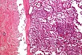
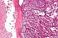
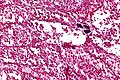
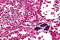
| ===Microscopic===
| | ==Microscopic== |
| Features:<ref name=Ref_WMSP627>{{Ref WMSP|627}}</ref> | | Features:<ref name=Ref_WMSP627>{{Ref WMSP|627}}</ref> |
| *Arranged in nest/separated by thin septa; vaguely resembles alveoli (at low power). | | *Arranged in nests/separated by thin septa; vaguely resembles alveoli (at low power). |
| *Large cells (~30-50 μm) with abundant eosinophilic cytoplasm. | | *Large cells (~30-50 μm) with abundant eosinophilic cytoplasm. |
| | **May be focally cleared. |
| *An eccentric nucleus. | | *An eccentric nucleus. |
| *+/-Nucleolus. | | *+/-Nucleolus, prominent. |
| | *+/-Multi-nucleation (common). |
|
| |
|
| Images:
| | DDx: |
| *[http://commons.wikimedia.org/wiki/File:Alveolar_soft_part_sarcoma_-_low_mag.jpg ASPS - low mag. (WC)]. | | *[[Paraganglioma]]. |
| *[http://commons.wikimedia.org/wiki/File:Alveolar_soft_part_sarcoma_-_intermed_mag.jpg ASPS - intermed. mag. (WC)]. | | *[[Clear cell renal cell carcinoma]] - areas with cytoplasmic clearing. |
| *[http://commons.wikimedia.org/wiki/File:Alveolar_soft_part_sarcoma_-2-_very_high_mag.jpg ASPS - very high mag. (WC)]. | | *[[Rhabdomyosarcoma]], alveolar - usu. does not have sheets of rhabdoid cells, no cross striations. |
|
| |
|
| ===Molecular=== | | ===Images=== |
| *t(X;17)(p11.2;q25).<ref>{{OMIM|606243}}</ref> | | <gallery> |
| | Image:Alveolar soft part sarcoma - very low mag.jpg | ASPS - very low mag. (WC/Nephron) |
| | Image:Alveolar_soft_part_sarcoma_-_low_mag.jpg | ASPS - low mag. (WC/Nephron) |
| | Image:Alveolar_soft_part_sarcoma_-_intermed_mag.jpg | ASPS - intermed. mag. (WC/Nephron) |
| | Image:Alveolar soft part sarcoma - high mag.jpg | ASPS - high mag. (WC/Nephron) |
| | Image:Alveolar soft part sarcoma - very high mag.jpg | ASPS - very high mag. (WC/Nephron) |
| | Image:Alveolar_soft_part_sarcoma_-2-_very_high_mag.jpg | ASPS - very high mag. (WC/Nephron) |
| | </gallery> |
| | www: |
| | *[http://www.webpathology.com/image.asp?case=508&n=10 ASPS - PAS (webpathology.com)]. |
| | *[http://www.ncbi.nlm.nih.gov/pmc/articles/PMC1860509/figure/fig1/ ASPS - low mag. (nih.gov)].<ref name=pmid17071801/> |
| | *[http://www.ncbi.nlm.nih.gov/pmc/articles/PMC1860509/figure/fig5/ ASPS - high mag. (nih.gov)].<ref name=pmid17071801/> |
|
| |
|
| ==Desmoplastic small round cell tumour== | | ==Stains== |
| *Abbreviated ''DSRCT''. | | *PAS +ve (cytoplasmic) - considered the most useful.<ref name=pmid17516754>{{Cite journal | last1 = Zarrin-Khameh | first1 = N. | last2 = Kaye | first2 = KS. | title = Alveolar soft part sarcoma. | journal = Arch Pathol Lab Med | volume = 131 | issue = 3 | pages = 488-91 | month = Mar | year = 2007 | doi = 10.1043/1543-2165(2007)131[488:ASPS]2.0.CO;2 | PMID = 17516754 }}</ref> |
| ===General=== | | *PASD +ve (cytoplasmic). |
| *Males > females.
| |
| *Usu. affects young adults.
| |
| *Typically retroperitoneal.
| |
| *Poor prognosis. | |
|
| |
|
| ===Microscopic=== | | ==IHC== |
| Features:<ref name=pmid10207460>{{cite journal |author=Pickhardt PJ, Fisher AJ, Balfe DM, Dehner LP, Huettner PC |title=Desmoplastic small round cell tumor of the abdomen: radiologic-histopathologic correlation |journal=Radiology |volume=210 |issue=3 |pages=633–8 |year=1999 |month=March |pmid=10207460 |doi= |url=http://radiology.rsna.org/content/210/3/633.full}}</ref>
| | *TFE3 +ve -- suggestive of characteristic translocation. |
| #Broad bands of paucicellular fibrous stroma with:
| | *Desmin ~ 50% of cases.<ref name=pmid17071801/> |
| #Small round cells in nests with an undulating sharp border.
| |
| #*The small round cells lack distinct nucleoli and have scant cytoplasm; they are ''[[small round cell tumour]]'' cells.
| |
|
| |
|
| Notes:
| | Others:<ref name=pmid17071801/> |
| *Usu. abundant mitoses. | | *EMA -ve. |
| *+/-Necrosis. | | *Cytokeratins -ve. |
| | *HMB45 -ve. |
| | *Melan‐A -ve. |
| | *Chromogranin A -ve. |
| | *Synaptophysin -ve. |
|
| |
|
| Images:
| | ==Molecular== |
| *[http://en.wikipedia.org/wiki/File:Desmoplastic_small_round_cell_tumour_-_intermed_mag.jpg DSRCT - intermed. mag. (WC)].
| | *t(X;17)(p11.2;q25).<ref>{{OMIM|606243}}</ref> |
| *[http://en.wikipedia.org/wiki/File:Desmoplastic_small_round_cell_tumour_-_very_high_mag.jpg DSRCT - very high mag. (WC)].
| |
| | |
| DDx:
| |
| *Metastatic [[germ cell tumour]] (DDx of location and age).
| |
| *[[Embryonal RMS]].
| |
| **It should be noted that DSRCT, like embryonal RMS, is +ve for desmin!
| |
| | |
| ===IHC===
| |
| Features:
| |
| *AE1/AE3 +ve.
| |
| *Desmin +ve.
| |
| *EMA +ve.
| |
| | |
| ===Molecular===
| |
| *t(11;22)(p13;q12).<ref name=pmid17964965>{{cite journal |author=Lee YS, Hsiao CH |title=Desmoplastic small round cell tumor: a clinicopathologic, immunohistochemical and molecular study of four patients |journal=J. Formos. Med. Assoc. |volume=106 |issue=10 |pages=854–60 |year=2007 |month=October |pmid=17964965 |doi=10.1016/S0929-6646(08)60051-0 |url=}}</ref><ref>{{cite journal |author=Lal DR, Su WT, Wolden SL, Loh KC, Modak S, La Quaglia MP |title=Results of multimodal treatment for desmoplastic small round cell tumors |journal=J. Pediatr. Surg. |volume=40 |issue=1 |pages=251–5 |year=2005 |month=January |pmid=15868593 |doi=10.1016/j.jpedsurg.2004.09.046 |url=http://www.dsrct.com/JPS%20Article.pdf}}</ref> | |
| | |
| ==Clear cell sarcoma==
| |
| *Known among pathologists as "soft-tissue melanoma" and "melanoma of the soft parts", as it has a strong morphological resemblance.<ref name=pmid18300804>{{cite journal |author=Hisaoka M, Ishida T, Kuo TT, ''et al.'' |title=Clear cell sarcoma of soft tissue: a clinicopathologic, immunohistochemical, and molecular analysis of 33 cases |journal=Am. J. Surg. Pathol. |volume=32 |issue=3 |pages=452–60 |year=2008 |month=March |pmid=18300804 |doi=10.1097/PAS.0b013e31814b18fb |url=}}</ref>
| |
| **Molecular changes and origin distinct from melanoma.
| |
| *Incidence: rare soft tissue tumour.
| |
|
| |
|
| ===Clinical===
| | Note: |
| *Usually - deep soft tissue ''or'' extremities. | | *Same translocation may be seen in ''[[renal tumour with Xp11.2 translocation]]''. |
| *Guarded prognosis.
| |
| *First described in 1965.<ref>URL: [http://www.informaworld.com/smpp/723576818-750600/ftinterface~db=all~content=a789166263~fulltext=713240928 http://www.informaworld.com/smpp/723576818-750600/ftinterface~db=all~content=a789166263~fulltext=713240928]. Accessed on: 5 May 2010.</ref>
| |
|
| |
|
| ===Microscopy=== | | ==EM== |
| Features:<ref name=pmid18300804/>
| | *Distinctive membrane-bound intracytoplasmic crystal lattice with ~5 nm fibres spaced 10 nm apart.<ref name=pmid17071801/> |
| *Architecture: sheets or fascicular (bundles) arrangement.
| |
| *Cells: Spindle cells or epithelioid cells.
| |
| *Prominent nucleoli - basophilic.
| |
| *Fibrous septae.
| |
| *Uniform
| |
|
| |
|
| Image: | | Image: |
| *[http://commons.wikimedia.org/wiki/File:Clear_cell_sarcoma.Image12.jpg Clear cell sarcoma (WC)]. | | *[http://www.ncbi.nlm.nih.gov/pmc/articles/PMC1860509/figure/fig8/ Distinctive crystal lattice in ASPS (nlm.nih.gov)].<ref name=pmid17071801/> |
| *[http://www.informaworld.com/ampp/image?path=/713690780/789166263/sonc_a_284443_o_f0003g.jpeg Clear cell sarcoma (informaworld.com)].
| |
| | |
| ===IHC===
| |
| Features:<ref name=pmid18300804/>
| |
| *S100 +ve.
| |
| *HMB-45 +ve.
| |
| *Melan A (MART-1) +ve; sometimes -ve.
| |
| *BCL2 +ve.
| |
| *CD57 +ve (usually).
| |
| | |
| Keratins:
| |
| *EMA may be +ve.
| |
| *CAM5.2 -ve.
| |
| *AE1/AE3 -ve.
| |
| | |
| ===Molecular studies===
| |
| *Chromosomal translocation t(12;22)(q13;q12).<ref name=pmid18300804/>
| |
| **Fusion transcripts:
| |
| ***EWSR1-ATF1.
| |
| ***EWSR1-CREB1 (GI tract associated).
| |
| | |
| ==Synovial sarcoma==
| |
| ===General===
| |
| *Does not arise from cartilage.<ref name=Ref_WMSP627>{{Ref WMSP|627}}</ref>
| |
| *Young adults or adolescents.
| |
| | |
| ===Microscopic===
| |
| Comes in three flavours:<ref name=Ref_WMSP627>{{Ref WMSP|627}}</ref><ref>{{cite journal |author=Schaal CH, Navarro FC, Moraes Neto FA |title=Primary renal sarcoma with morphologic and immunohistochemical aspects compatible with synovial sarcoma |journal=Int Braz J Urol |volume=30 |issue=3 |pages=210–3 |year=2004 |pmid=15689250 |doi= |url=http://www.brazjurol.com.br/may_june_2004/Schaal_ing_210_213.htm}}</ref>
| |
| #Spindle cell sarcoma with features of hemangiopericytoma, i.e. staghorn vessels.
| |
| #Biphasic synovial sarcoma:
| |
| ##Spindle cells with features of hemangiopericytoma.
| |
| ##Epitheliod glands or nests.
| |
| #Primative round cell type.
| |
| | |
| Images:
| |
| *[http://www.scielo.br/img/revistas/ibju/v30n3/3a06f03.jpg Synovial sarcoma (scielo.br)].
| |
| *[http://www.humpath.com/spip.php?page=article&id_article=1965 Synovial sarcoma - collection of images (humpath.com)].
| |
| | |
| ===IHC===
| |
| Features:<ref name=Ref_WMSP627>{{Ref WMSP|627}}</ref>
| |
| *Vimentin +ve + cytokeratin and/or EMA +ve.
| |
| *CD99 +ve.
| |
| | |
| Others:
| |
| *Beta-catenin +ve ~30-70%.<ref name=pmid16740029>{{cite journal |author=Horvai AE, Kramer MJ, O'Donnell R |title=Beta-catenin nuclear expression correlates with cyclin D1 expression in primary and metastatic synovial sarcoma: a tissue microarray study |journal=Arch. Pathol. Lab. Med. |volume=130 |issue=6 |pages=792–8 |year=2006 |month=June |pmid=16740029 |doi= |url=}}</ref>
| |
| *Cyclin D1 ~50%.<ref name=pmid16740029/><ref name=pmid15375433>{{cite journal |author=Ng TL, Gown AM, Barry TS, ''et al.'' |title=Nuclear beta-catenin in mesenchymal tumors |journal=Mod. Pathol. |volume=18 |issue=1 |pages=68–74 |year=2005 |month=January |pmid=15375433 |doi=10.1038/modpathol.3800272 |url=}}</ref>
| |
| | |
| ===Molecular pathology===
| |
| Unique translocation:
| |
| *t(X;18)(p11.2;q11.2).<ref>URL: [http://www.ncbi.nlm.nih.gov/omim/300813 http://www.ncbi.nlm.nih.gov/omim/300813]. Accessed on: 30 May 2010.</ref>
| |
|
| |
|
| =See also= | | ==See also== |
| *[[Bone]]. | | *[[Soft tissue lesions]]. |
| *[[Dermatopathology]].
| | *[[Head and neck pathology]]. |
| *[[Hematopathology]].
| |
| *[[Spindle cell lesion]].
| |
| *[[Neurofibromatosis]].
| |
| *[[Small round cell tumours]]. | |
|
| |
|
| =References= | | ==References== |
| {{reflist|2}} | | {{Reflist|2}} |
|
| |
|
| [[Category:Soft tissue lesions]] | | [[Category:Soft tissue lesions]] |
| | [[Category:Diagnosis]] |