Difference between revisions of "Pheochromocytoma"
(+cat.) |
(split out) |
||
| Line 1: | Line 1: | ||
# | '''Pheochromocytoma''' is a tumour of the [[adrenal gland]] medulla. It may be benign of malignant. | ||
==General== | |||
*Considered to be a [[paraganglioma]].<ref name=Ref_EP327>{{Ref EP|327}}</ref> | |||
*Literally means "dusky" (pheo) "colour" (chromo) - dull appearance on gross. | |||
*Tumour arises from adrenal medulla - chromaffin cells.<ref name=Ref_PCPBoD8_586>{{Ref PCPBoD8|586}}</ref> | |||
Memory device - the rule of 10s:<ref name=Ref_PCPBoD8_586>{{Ref PCPBoD8|586}}</ref> | |||
*10% extra-adrenal (e.g. carotid body, organ of Zuckerkandl (neighourhood of aortic bifuration/IMA branch point)). | |||
*10% bilateral. | |||
*10% malignant. | |||
*10% no hypertension. | |||
*25% associated within a syndrome: | |||
*#[[Multiple endocrine neoplasia]] 2A and 2B. | |||
*#[[von Hippel-Lindau syndrome]]. | |||
*#[[Neurofibromatosis]] type 1. | |||
*#Familial paraganglioma syndromes - several. | |||
===Clinical=== | |||
*Classic finding: hypertension. | |||
*Paroxysms (i.e. episodes) of tachycardia, headache, anxiety, [[hypertension]]. | |||
Laboratory findings (urine): | |||
*Vanillylmandelic acid (VMA). | |||
*Metanephrines. | |||
==Microscopic== | |||
Features:<ref>{{Ref PBoD8|1161}}</ref> | |||
*Chief cells: | |||
**Usu. polygonal cells, may be spindled. | |||
**Arranged in cell nests - "Zellballen" (literally ''cell balls'') - '''key feature'''. | |||
**Stippled chromatin ([[AKA]] salt and pepper chromatin) - coarsely granular chromatin. | |||
**Granular cytoplasm, often basophilic - '''important'''. | |||
*Sustentacular cells (structural support cell). | |||
*Often haemorrhagic - highly vascular. | |||
*+/-Nuclear pleomorphism. | |||
Notes: | |||
*The nested architecture (Zellballen) is useful for differentiating from [[ACC]]. | |||
*[[Metastasis]] sole criteria of malignancy.<ref name=Ref_PCPBoD8_586>{{Ref PCPBoD8|586}}</ref> | |||
*Surrounding adrenal cortex is typically compressed.<ref>URL: [http://www.pathpedia.com/Education/eAtlas/Histopathology/Adrenal/Pheochromocytoma.aspx http://www.pathpedia.com/Education/eAtlas/Histopathology/Adrenal/Pheochromocytoma.aspx]. Accessed on: 27 May 2013.</ref> | |||
DDx: | |||
*[[Adrenal cortical carcinoma]] - ''[[pheochromocytoma versus adrenal cortical carcinoma]]''. | |||
===Images=== | |||
<gallery> | |||
Image:Carotid_body_tumour_2_low_mag.jpg | Carotid body tumour - low mag. (WC/Nephron) | |||
Image:Carotid_body_tumour_2_high_mag.jpg | Carotid body tumour - high mag. (WC/Nephron) | |||
</gallery> | |||
====Pheochromocytoma versus adrenal cortical carcinoma==== | |||
*Pheochromocytoma and adrenal cortical carcinoma overlap histologically.<ref name=pmid20154585>{{Cite journal | last1 = Sangoi | first1 = AR. | last2 = McKenney | first2 = JK. | title = A tissue microarray-based comparative analysis of novel and traditional immunohistochemical markers in the distinction between adrenal cortical lesions and pheochromocytoma. | journal = Am J Surg Pathol | volume = 34 | issue = 3 | pages = 423-32 | month = Mar | year = 2010 | doi = 10.1097/PAS.0b013e3181cfb506 | PMID = 20154585 }}</ref> | |||
Favour pheochromocytoma: | |||
*Small chickenwire-pattern blood vessels, nests, salt-and-pepper chromatin, red blood cell extravasation. | |||
Favour adrenal cortical carcinoma: | |||
*Nucleolus, sheeting. | |||
===Malignant pheochromoctyoma=== | |||
#''Robbins'' says metastases are the sole criteria of malignancy.<ref name=Ref_PCPBoD8_586>{{Ref PCPBoD8|586}}</ref> | |||
#''Thompson'' suggests one can differentiate benign from malignant with the aid of the following:<ref name=Ref_EP259>{{Ref EP|259}}</ref> | |||
#*Marked nuclear atypia. | |||
#*Invasion: | |||
#**Capsular. | |||
#**Vascular. | |||
#*Necrosis. | |||
#*Cellular monotony. | |||
#*Mitoses: | |||
#**Rate. | |||
#**Atypical mitosis. | |||
==IHC== | |||
*Chief cells: | |||
**Chromogranin A +ve. | |||
**Synaptophysin +ve. | |||
*Sustentacular cells: | |||
**S100 +ve. | |||
==[[Electron microscopy]]== | |||
*Membrane-bound secretory granules. | |||
==Sign out== | |||
<pre> | |||
ADRENAL MASS, RIGHT, ADRENALECTOMY: | |||
- PHEOCHROMOCYTOMA. | |||
- SURGICAL MARGIN NEGATIVE FOR PHEOCHROMOCYTOMA. | |||
COMMENT: | |||
The tumour cells stains for chromogranin and synaptophysin. S-100 marks the sustentacular cells. | |||
Inhibin is negative in the tumour cells. The immunostaining pattern is consistent with a | |||
pheochromocytoma. | |||
</pre> | |||
====Micro==== | |||
The sections shows a partially hemorrhagic lesion in the medulla of the adrenal gland that is arranged in nests (Zellballen). The tumour cells have abundant grey/blue granular cytoplasm, and nuclei with granular chromatin (salt and pepper chromatin). The lesion is surrounded by a compressed rim of adrenal cortex and fibrosis tissue. The core of the lesion is fibrotic and has clusters of hemosiderin-laden macrophages. | |||
There is no capsular invasion. Vascular invasion is not identified. There is no necrosis. Mitotic activity is not appreciated. | |||
The adrenal cortex is unremarkable. | |||
==See also== | |||
*[[Paraganglioma]]. | |||
*[[Adrenal gland]]. | |||
==References== | |||
{{Reflist|2}} | |||
[[Category:Diagnosis]] | [[Category:Diagnosis]] | ||
[[Category:Adrenal gland]] | |||
Revision as of 02:49, 23 August 2014
Pheochromocytoma is a tumour of the adrenal gland medulla. It may be benign of malignant.
General
- Considered to be a paraganglioma.[1]
- Literally means "dusky" (pheo) "colour" (chromo) - dull appearance on gross.
- Tumour arises from adrenal medulla - chromaffin cells.[2]
Memory device - the rule of 10s:[2]
- 10% extra-adrenal (e.g. carotid body, organ of Zuckerkandl (neighourhood of aortic bifuration/IMA branch point)).
- 10% bilateral.
- 10% malignant.
- 10% no hypertension.
- 25% associated within a syndrome:
- Multiple endocrine neoplasia 2A and 2B.
- von Hippel-Lindau syndrome.
- Neurofibromatosis type 1.
- Familial paraganglioma syndromes - several.
Clinical
- Classic finding: hypertension.
- Paroxysms (i.e. episodes) of tachycardia, headache, anxiety, hypertension.
Laboratory findings (urine):
- Vanillylmandelic acid (VMA).
- Metanephrines.
Microscopic
Features:[3]
- Chief cells:
- Usu. polygonal cells, may be spindled.
- Arranged in cell nests - "Zellballen" (literally cell balls) - key feature.
- Stippled chromatin (AKA salt and pepper chromatin) - coarsely granular chromatin.
- Granular cytoplasm, often basophilic - important.
- Sustentacular cells (structural support cell).
- Often haemorrhagic - highly vascular.
- +/-Nuclear pleomorphism.
Notes:
- The nested architecture (Zellballen) is useful for differentiating from ACC.
- Metastasis sole criteria of malignancy.[2]
- Surrounding adrenal cortex is typically compressed.[4]
DDx:
Images
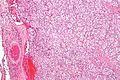
Carotid body tumour - low mag. (WC/Nephron)
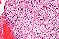
Carotid body tumour - high mag. (WC/Nephron)


Pheochromocytoma versus adrenal cortical carcinoma
- Pheochromocytoma and adrenal cortical carcinoma overlap histologically.[5]
Favour pheochromocytoma:
- Small chickenwire-pattern blood vessels, nests, salt-and-pepper chromatin, red blood cell extravasation.
Favour adrenal cortical carcinoma:
- Nucleolus, sheeting.
Malignant pheochromoctyoma
- Robbins says metastases are the sole criteria of malignancy.[2]
- Thompson suggests one can differentiate benign from malignant with the aid of the following:[6]
- Marked nuclear atypia.
- Invasion:
- Capsular.
- Vascular.
- Necrosis.
- Cellular monotony.
- Mitoses:
- Rate.
- Atypical mitosis.
IHC
- Chief cells:
- Chromogranin A +ve.
- Synaptophysin +ve.
- Sustentacular cells:
- S100 +ve.
Electron microscopy
- Membrane-bound secretory granules.
Sign out
ADRENAL MASS, RIGHT, ADRENALECTOMY:
- PHEOCHROMOCYTOMA.
- SURGICAL MARGIN NEGATIVE FOR PHEOCHROMOCYTOMA.

COMMENT:
The tumour cells stains for chromogranin and synaptophysin. S-100 marks the sustentacular cells.
Inhibin is negative in the tumour cells. The immunostaining pattern is consistent with a
pheochromocytoma.
Micro
The sections shows a partially hemorrhagic lesion in the medulla of the adrenal gland that is arranged in nests (Zellballen). The tumour cells have abundant grey/blue granular cytoplasm, and nuclei with granular chromatin (salt and pepper chromatin). The lesion is surrounded by a compressed rim of adrenal cortex and fibrosis tissue. The core of the lesion is fibrotic and has clusters of hemosiderin-laden macrophages.
There is no capsular invasion. Vascular invasion is not identified. There is no necrosis. Mitotic activity is not appreciated.
The adrenal cortex is unremarkable.
See also
References
- ↑ Thompson, Lester D. R. (2006). Endocrine Pathology: A Volume in Foundations in Diagnostic Pathology Series (1st ed.). Churchill Livingstone. pp. 327. ISBN 978-0443066856.
- ↑ 2.0 2.1 2.2 2.3 Mitchell, Richard; Kumar, Vinay; Fausto, Nelson; Abbas, Abul K.; Aster, Jon (2011). Pocket Companion to Robbins & Cotran Pathologic Basis of Disease (8th ed.). Elsevier Saunders. pp. 586. ISBN 978-1416054542.
- ↑ Kumar, Vinay; Abbas, Abul K.; Fausto, Nelson; Aster, Jon (2009). Robbins and Cotran pathologic basis of disease (8th ed.). Elsevier Saunders. pp. 1161. ISBN 978-1416031215.
- ↑ URL: http://www.pathpedia.com/Education/eAtlas/Histopathology/Adrenal/Pheochromocytoma.aspx. Accessed on: 27 May 2013.
- ↑ Sangoi, AR.; McKenney, JK. (Mar 2010). "A tissue microarray-based comparative analysis of novel and traditional immunohistochemical markers in the distinction between adrenal cortical lesions and pheochromocytoma.". Am J Surg Pathol 34 (3): 423-32. doi:10.1097/PAS.0b013e3181cfb506. PMID 20154585.
- ↑ Thompson, Lester D. R. (2006). Endocrine Pathology: A Volume in Foundations in Diagnostic Pathology Series (1st ed.). Churchill Livingstone. pp. 259. ISBN 978-0443066856.