Difference between revisions of "Angiomyofibroblastoma"
Jump to navigation
Jump to search
(+images) |
|||
| Line 1: | Line 1: | ||
{{ Infobox diagnosis | {{ Infobox diagnosis | ||
| Name = {{PAGENAME}} | | Name = {{PAGENAME}} | ||
| Image = | | Image = Angiomyofibroblastoma humpath 4.jpg | ||
| Width = | | Width = | ||
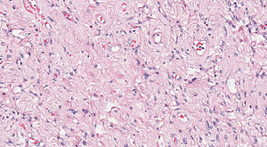
| Caption = | | Caption = Angiomyofibroblastoma. [[H&E stain]]. | ||
| Synonyms = | | Synonyms = | ||
| Micro = hypercellular zones and hypocellular edematous zones, small blood vessels (~20 micrometers) - no large blood vessels, [[myxoid stroma]], small stellate cell/spindle cells without significant nuclear atypia | | Micro = hypercellular zones and hypocellular edematous zones, small blood vessels (~20 micrometers) - no large blood vessels, [[myxoid stroma]], small stellate cell/spindle cells without significant nuclear atypia | ||
| Line 59: | Line 59: | ||
===Images=== | ===Images=== | ||
<gallery> | |||
Image:Angiomyofibroblastoma humpath 1.jpg | AMF - low mag. | |||
Image:Angiomyofibroblastoma humpath 2.jpg | AMF - intermed. mag. | |||
Image:Angiomyofibroblastoma humpath 3.jpg | AMF - high mag. | |||
Image:Angiomyofibroblastoma humpath 4.jpg | AMF - very high mag. | |||
</gallery> | |||
www: | www: | ||
*[http://www.webpathology.com/image.asp?case=544&n=4 Angiomyofibroblastoma (webpathology.com)] | *[http://www.webpathology.com/image.asp?case=544&n=4 Angiomyofibroblastoma (webpathology.com)] | ||
Latest revision as of 07:56, 8 May 2014
| Angiomyofibroblastoma | |
|---|---|
| Diagnosis in short | |
 Angiomyofibroblastoma. H&E stain. | |
|
| |
| LM | hypercellular zones and hypocellular edematous zones, small blood vessels (~20 micrometers) - no large blood vessels, myxoid stroma, small stellate cell/spindle cells without significant nuclear atypia |
| LM DDx | aggressive angiomyxoma |
| Gross | well-circumscribed |
| Site | female pelvis (vulva), case reports of similar lesion in males |
|
| |
| Clinical history | slow growing, usu. not painful |
| Prevalence | very rare |
| Clin. DDx | Bartholin cyst, aggressive angiomyxoma |
Angiomyofibroblastoma, abbreviated AMF,[1] is rare uncommon soft tissue lesion of the vulva.
General
Clinical:[4]
- Slow growing.
- Uncommonly recur.
- Usually painless.
Clinical DDx:
Gross
- Vulvar lesion.
- Well-circumscribed.[4]
Microscopic
Features:[5]
- Hypercellular zones and hypocellular edematous zones.
- Small blood vessels (~20 micrometers) - no large blood vessels - key feature.
- Myxoid stroma - key feature.
- Small stellate cell/spindle cells without significant nuclear atypia.
DDx:
- Aggressive angiomyxoma - less cellular, large blood vessels.
Images
www:
- Angiomyofibroblastoma (webpathology.com)
- Angiomyofibroblastoma - high mag. (webpathology.com).
- Angiomyofibroblastoma-like tumour of scrotum (nih.gov).[2]
IHC
See also
References
- ↑ Cao D, Srodon M, Montgomery EA, Kurman RJ (April 2005). "Lipomatous variant of angiomyofibroblastoma: report of two cases and review of the literature". Int. J. Gynecol. Pathol. 24 (2): 196–200. PMID 15782077.
- ↑ 2.0 2.1 Ding, G.; Yu, Y.; Jin, M.; Xu, J.; Zhang, Z. (Feb 2014). "Angiomyofibroblastoma-like tumor of the scrotum: A case report and literature review.". Oncol Lett 7 (2): 435-438. doi:10.3892/ol.2013.1741. PMID 24396463.
- ↑ Butt, Z.; Slatnik, J.; Estey, EP. (Dec 2013). "Robotic assisted laparoscopic excision of a pelvic angiomyofibroblastoma-like tumor.". Can J Urol 20 (6): 7067-9. PMID 24331351.
- ↑ 4.0 4.1 4.2 4.3 Seo, JW.; Lee, KA.; Yoon, NR.; Lee, JW.; Kim, BG.; Bae, DS. (Sep 2013). "Angiomyofibroblastoma of the vulva.". Obstet Gynecol Sci 56 (5): 349-51. doi:10.5468/ogs.2013.56.5.349. PMID 24328028.
- ↑ Fletcher, CD.; Tsang, WY.; Fisher, C.; Lee, KC.; Chan, JK. (Apr 1992). "Angiomyofibroblastoma of the vulva. A benign neoplasm distinct from aggressive angiomyxoma.". Am J Surg Pathol 16 (4): 373-82. PMID 1314521.


