Difference between revisions of "Pleomorphic adenoma"
Jump to navigation
Jump to search
(split-out) |
m |
||
| Line 24: | Line 24: | ||
*Proliferation of myoepithelium and epithelium (ductal cells) in mesenchymal stroma. | *Proliferation of myoepithelium and epithelium (ductal cells) in mesenchymal stroma. | ||
**Cells in ducts = epithelial. | **Cells in ducts = epithelial. | ||
**Cells not in ducts = myoepithelial.<ref name=IW_10jan2011> | **Cells not in ducts = myoepithelial.<ref name=IW_10jan2011>Weinreb I. 10 January 2011.</ref> | ||
*Mesenchymal stroma - '''important feature'''. | *Mesenchymal stroma - '''important feature'''. | ||
**May be any of following: [[myxoid stroma|myxoid]], mucochondroid, hyalinized, osseous, fatty. | **May be any of following: [[myxoid stroma|myxoid]], mucochondroid, hyalinized, osseous, fatty. | ||
| Line 31: | Line 31: | ||
Notes: | Notes: | ||
*Mesenchymal stroma not required for diagnosis -- if >5% ducts.<ref name=IW_10jan2011> | *Mesenchymal stroma not required for diagnosis -- if >5% ducts.<ref name=IW_10jan2011>Weinreb I. 10 January 2011.</ref> | ||
**No chondroid stroma ''and'' <5% ductal cells = '''[[myoepithelioma]]'''. | **No chondroid stroma ''and'' <5% ductal cells = '''[[myoepithelioma]]'''. | ||
*Complete excision is often elusive; stating "completely excised" on a surgical pathology report is unwise. | *Complete excision is often elusive; stating "completely excised" on a surgical pathology report is unwise. | ||
Revision as of 15:29, 10 November 2013
Pleomorphic adenoma, abbreviated PA, is a very common benign salivary gland tumour.
General
Features:
- Very common - approx. 60% of parotid gland tumours.[1]
- May transform into a malignant tumour.
- Other benign salivary gland tumours do not do this.
- Only benign childhood salivary gland tumour of significance.
Weinreb's dictums
- Most common salivary tumour in all age groups.
- Seen in all sites (unlike other benign tumours).
- Recurrence and malignancy risk (unlike other benign salivary gland tumours).
- Any part of a tumour that looks like PA makes it a PA.
Gross
- May be cartilaginous appearing.
Image:
Microscopic
Features:[1]
- Proliferation of myoepithelium and epithelium (ductal cells) in mesenchymal stroma.
- Cells in ducts = epithelial.
- Cells not in ducts = myoepithelial.[2]
- Mesenchymal stroma - important feature.
Notes:
- Mesenchymal stroma not required for diagnosis -- if >5% ducts.[2]
- No chondroid stroma and <5% ductal cells = myoepithelioma.
- Complete excision is often elusive; stating "completely excised" on a surgical pathology report is unwise.
- Look for, i.e. rule-out, poorly differentiated carcinoma: carcinoma ex pleomorphic adenoma.
Memory device: MEC = myoepithelium, epithelium, chondromyxoid stroma.
DDx:
Images
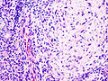
PA. (WC)
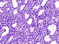
PA. (WC)
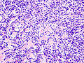
PA. (WC)
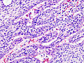
PA. (WC)
_parotid_gland.jpg)
_parotid_gland.jpg)
_parotid_gland.jpg)
_parotid_gland.jpg)
www:
IHC
- S-100 +ve, SMA +ve, GFAP +ve.