Difference between revisions of "Rheumatoid arthritis"
Jump to navigation
Jump to search
(→Rheumatoid nodule: +images) |
(→Joints: tweak) |
||
| Line 10: | Line 10: | ||
*[[Neutrophil]]s. | *[[Neutrophil]]s. | ||
== | ==Joint with rheumatic disease== | ||
{{Main|Joints}} | {{Main|Joints}} | ||
*[[AKA]] ''rheumatic joint disease''. | |||
===General=== | |||
Clinical: | |||
*Tumour - swelling. | |||
*Rubor - redness. | |||
*Calor - heat. | |||
*Dolor - pain. | |||
===Microscopic=== | ===Microscopic=== | ||
Features: | Features:<ref name=Ref_WMSP660>{{Ref WMSP|660}}</ref> | ||
*Chronic inflammation | *Chronic inflammation, esp. lymphocytes. | ||
**+/-Lymphoid follicles. | |||
*Synovial hyperplasia - with papillary ''or'' polyoid architecture. | |||
**Synoviocytes may show binucleation and mild atypia. | |||
*+/-Fibrin. | |||
*+/-Bone. | |||
*+/-Cartilage. | |||
Note: | |||
*Changes are non-specific - DDx includes other rheumatic diseases ([[systemic lupus erythematosus]], [[psoriatic arthritis]]). | |||
Images: | Images: | ||
Revision as of 16:17, 29 October 2012
Rheumatoid arthritis, commonly abbreviated RA, is an autoimmune disorder.
Skin
General
- May manifest as rheumatoid neutrophilic dermatitis - super rare.[1]
Microscopic
Features:
- Nodular and diffuse pattern.
- Neutrophils.
Joint with rheumatic disease
Main article: Joints
- AKA rheumatic joint disease.
General
Clinical:
- Tumour - swelling.
- Rubor - redness.
- Calor - heat.
- Dolor - pain.
Microscopic
Features:[2]
- Chronic inflammation, esp. lymphocytes.
- +/-Lymphoid follicles.
- Synovial hyperplasia - with papillary or polyoid architecture.
- Synoviocytes may show binucleation and mild atypia.
- +/-Fibrin.
- +/-Bone.
- +/-Cartilage.
Note:
- Changes are non-specific - DDx includes other rheumatic diseases (systemic lupus erythematosus, psoriatic arthritis).
Images:
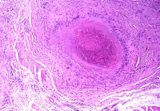
Rheumatoid nodule
General
- Usually only in seropositive cases.[4]
Microscopic
- Necrotic collagen bundles with fibrin surrounded by:
- Palisading granuloma.
- +/-Eosinophils.
Notes:
- Histomorphologically very similar to Granuloma annulare.
DDx:
- Granuloma annulare - has mucin in the core of the granuloma.[5]
- Necrobiosis lipoidica.
Images:
- www:
- WC:
Pleural disease
- See Rheumatoid pleuritis.
Lung disease
- See Medical lung disease.
RA may involve the lung.
Amyloidosis
- See Amyloidosis.
Amyloidosis may be due to RA.
Felty syndrome
RA may occur in Felty syndrome -- the triad:[8]
- Rheumatoid arthritis.
- Splenomegaly.
- Neutropenia.
Felty syndrome is associated with large granular lymphocytic leukemia.[8][9]
See also
References
- ↑ Mashek, HA.; Pham, CT.; Helm, TN.; Klaus, M. (Jun 1997). "Rheumatoid neutrophilic dermatitis.". Arch Dermatol 133 (6): 757-60. PMID 9197831.
- ↑ Humphrey, Peter A; Dehner, Louis P; Pfeifer, John D (2008). The Washington Manual of Surgical Pathology (1st ed.). Lippincott Williams & Wilkins. pp. 660. ISBN 978-0781765275.
- ↑ URL: http://library.med.utah.edu/WebPath/EXAM/IMGQUIZ/msfrm.html. Accessed on: 5 December 2010.
- ↑ 4.0 4.1 Tadrous, Paul.J. Diagnostic Criteria Handbook in Histopathology: A Surgical Pathology Vade Mecum (1st ed.). Wiley. pp. 299. ISBN 978-0470519035.
- ↑ 5.0 5.1 Busam, Klaus J. (2009). Dermatopathology: A Volume in the Foundations in Diagnostic Pathology Series (1st ed.). Saunders. pp. 52. ISBN 978-0443066542.
- ↑ 6.0 6.1 URL: http://granuloma.homestead.com/palisading.html. Accessed on: 1 November 2010.
- ↑ URL: http://www.pathguy.com/lectures/joints.htm. Accessed on: 1 November 2010.
- ↑ 8.0 8.1 Mitchell, Richard; Kumar, Vinay; Fausto, Nelson; Abbas, Abul K.; Aster, Jon (2011). Pocket Companion to Robbins & Cotran Pathologic Basis of Disease (8th ed.). Elsevier Saunders. pp. 328. ISBN 978-1416054542.
- ↑ Liu, X.; Loughran, TP. (Jul 2011). "The spectrum of large granular lymphocyte leukemia and Felty's syndrome.". Curr Opin Hematol 18 (4): 254-9. doi:10.1097/MOH.0b013e32834760fb. PMID 21546829.