Intestinal metaplasia of the stomach
(Redirected from Stomach IM)
Jump to navigation
Jump to search
| Intestinal metaplasia of the stomach | |
|---|---|
| Diagnosis in short | |
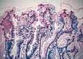
 Intestinal metaplasia of the stomach. H&E stain. | |
|
| |
| LM | goblet cells with foveolar epithelium |
| LM DDx | contaminant from the duodenum, gastric dysplasia, gastric carcinoma, gastric heterotopia of the duodenum |
| Stains | alcian blue/PAS +ve |
| IHC | CDX2 +ve |
| Site | stomach |
|
| |
| Associated Dx | gastric carcinoma, chronic gastritis, Helicobacter gastritis |
| Clinical history | +/-chronic gastritis |
| Endoscopy | +/-erythema |
| Clin. DDx | gastritis, gastric dysplasia |
| Intestinal metaplasia of the stomach | |
|---|---|
| External resources | |
| EHVSC | 10167 (focal) |
Intestinal metaplasia of the stomach is a relative common finding that is associated with a modest increased risk of gastric carcinoma.
It is also known as gastric intestinal metaplasia and may be abbreviated IM.
General
- Often part of surgical pathology report, e.g. "negative for intestinal metaplasia" or "intestinal metaplasia present".
- May be associated with Helicobacter spp. infection -- though Helicobacter don't like intestinal type mucosa, i.e. H. pylori are not typically found in regions with intestinal metaplasia.
- May be reversible - some epidemiological evidence.[1]
- Associated with gastric carcinomas of Lynch syndrome;[2] however, surveillance may not be worthwhile.[3]
Significance:
- Moderate risk increase for carcinoma; risk less than for Barrett's esophagus.[4]
- Odds ratio for corpus (~5.8x) higher than antrum (2.3x) when compared to individuals without IM.[5]
Gross/endoscopic
- +/-Erythema.
- Often associated with chronic gastritis.
Microscopic
Features:
- Goblet cells are present in the stomach - key feature.[6]
- In H&E sections the vacuole often stains light grey.
- Foveolar epithelium should be present in the same fragment.
- +/-Paneth cells - deep in the glands.[7]
- Very rarely present.
- Very uncommon in isolation.
Notes:
- Intestinal metaplasia (IM) is occasionally subdivided:[8]
- Complete IM = goblet cells and (intestinal) brush border.
- Incomplete IM = mucus vacuoles of various sizes, no (intestinal) brush border.
- Goblet cells metaplasia starts in the neck region of the gastric glands.[9]
DDx:
- Normal small bowel mucosa - no foveolar epithelium present, possibly a contaminant from a concurrent duodenal biopsy.
- Gastric dysplasia, intestinal type.
- Gastric heterotopia of the duodenum.
Images
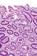
Stomach IM - intermed. mag. (WC)

Stomach IM - high mag. (WC)

Stains
- Alcian blue (pH 2.5)/PAS +ve.[10]
- Alican blue stain +ve.[citation needed]
Image
Barrett's mucosa - Alcian blue stain. (WC)

IHC
- CDX2 +ve (-ve in normal stomach).[1]
- Strong assoc. with Helicobacter gastritis as well as IM.[13]
Others:
- Lysozyme +ve - marks paneth cells.[7]
Sign out
Focal
Stomach, Antrum, Biopsy:
- Antral-type gastric mucosa with intestinal metaplasia (focal) and
  moderate chronic inactive inflammation.
- NEGATIVE for Helicobacter-like organisms.
- NEGATIVE for dysplasia and NEGATIVE for malignancy.
Block letters
STOMACH, BIOPSY:
- BODY-TYPE GASTRIC MUCOSA WITH INTESTINAL METAPLASIA, FOCAL.
- MINIMAL CHRONIC GASTRITIS (BODY OF STOMACH).
- NEGATIVE FOR HELICOBACTER-LIKE ORGANISMS.
- NEGATIVE FOR DYSPLASIA AND NEGATIVE FOR MALIGNANCY.
STOMACH, BIOPSY:
- ANTRAL-TYPE GASTRIC MUCOSA WITH FOCAL INTESTINAL METAPLASIA AND MILD CHRONIC
  INACTIVE INFLAMMATION.
- NEGATIVE FOR HELICOBACTER-LIKE ORGANISMS.
- NEGATIVE FOR DYSPLASIA AND NEGATIVE FOR MALIGNANCY.
Extensive
STOMACH, BIOPSY:
- SUPERFICIAL GASTRIC MUCOSA WITH EXTENSIVE INTESTINAL METAPLASIA AND MODERATE
  CHRONIC INACTIVE INFLAMMATION.
- NEGATIVE FOR HELICOBACTOR-LIKE ORGANISMS.
- NEGATIVE FOR DYSPLASIA AND NEGATIVE FOR MALIGNANCY.
See also
References
- ↑ 1.0 1.1 Walker, MM. (Jan 2003). "Is intestinal metaplasia of the stomach reversible?". Gut 52 (1): 1-4. PMC 1773527. PMID 12477745. https://www.ncbi.nlm.nih.gov/pmc/articles/PMC1773527/.
- ↑ Cristofaro, G.; Lynch, HT.; Caruso, ML.; Attolini, A.; DiMatteo, G.; Giorgio, P.; Senatore, S.; Argentieri, A. et al. (Jul 1987). "New phenotypic aspects in a family with Lynch syndrome II.". Cancer 60 (1): 51-8. PMID 3581033.
- ↑ Renkonen-Sinisalo, L.; Sipponen, P.; Aarnio, M.; Julkunen, R.; Aaltonen, LA.; Sarna, S.; Järvinen, HJ.; Mecklin, JP. (May 2002). "No support for endoscopic surveillance for gastric cancer in hereditary non-polyposis colorectal cancer.". Scand J Gastroenterol 37 (5): 574-7. PMID 12059060.
- ↑ Correa P, Piazuelo MB, Wilson KT (March 2010). "Pathology of gastric intestinal metaplasia: clinical implications". Am. J. Gastroenterol. 105 (3): 493–8. doi:10.1038/ajg.2009.728. PMC 2895407. PMID 20203636. http://www.ncbi.nlm.nih.gov/pmc/articles/PMC2895407/?tool=pubmed.
- ↑ Sakitani, K.; Hirata, Y.; Watabe, H.; Yamada, A.; Sugimoto, T.; Yamaji, Y.; Yoshida, H.; Maeda, S. et al. (Oct 2011). "Gastric cancer risk according to the distribution of intestinal metaplasia and neutrophil infiltration.". J Gastroenterol Hepatol 26 (10): 1570-5. doi:10.1111/j.1440-1746.2011.06767.x. PMID 21575058.
- ↑ URL: http://esynopsis.uchc.edu/eAtlas/GI/1280.htm. Accessed on: 16 August 2010.
- ↑ 7.0 7.1 Rubio, CA.; Befrits, R. (2009). "Increased lysozyme expression in gastric biopsies with intestinal metaplasia and pseudopyloric metaplasia.". Int J Clin Exp Med 2 (3): 248-53. PMID 19918317.
- ↑ Rugge, M.; Correa, P.; Dixon, MF.; Hattori, T.; Leandro, G.; Lewin, K.; Riddell, RH.; Sipponen, P. et al. (Feb 2000). "Gastric dysplasia: the Padova international classification.". Am J Surg Pathol 24 (2): 167-76. PMID 10680883.
- ↑ Stemmermann, GN.; Hayashi, T. (Sep 1968). "Intestinal metaplasia of the gastric mucosa: a gross and microscopic study of its distribution in various disease states.". J Natl Cancer Inst 41 (3): 627-34. PMID 4175677.
- ↑ Rivera-Hueto, F.; Lag-Asturiano, E.; Utrilla-Alcolea, JC.; Herrerías-Gutiérrez, JM. (Feb 2004). "Advanced gastric carcinoma with a complete intestinal metaplasia phenotype associated with early intestinal-type carcinoma.". Arch Pathol Lab Med 128 (2): 218-21. doi:10.1043/1543-2165(2004)128218:AGCWAC2.0.CO;2. PMID 14736279.
- ↑ Iida, F.; Kusama, J. (Dec 1982). "Gastric carcinoma and intestinal metaplasia. Significance of types of intestinal metaplasia upon development of gastric carcinoma.". Cancer 50 (12): 2854-8. PMID 7139576.
- ↑ Odze, Robert D.; Goldblum, John R. (2009). Surgical pathology of the GI tract, liver, biliary tract and pancreas (2nd ed.). Saunders. pp. 276. ISBN 978-1416040590.
- ↑ Satoh, K.; Mutoh, H.; Eda, A.; Yanaka, I.; Osawa, H.; Honda, S.; Kawata, H.; Kihira, K. et al. (Jun 2002). "Aberrant expression of CDX2 in the gastric mucosa with and without intestinal metaplasia: effect of eradication of Helicobacter pylori.". Helicobacter 7 (3): 192-8. PMID 12047325.