Solid pseudopapillary tumour
| Solid pseudopapillary tumour | |
|---|---|
| Diagnosis in short | |
 Solid pseudopapillary tumour. H&E stain. | |
|
| |
| Synonyms | solid pseudopapillary neoplasm, solid and papillary epithelial neoplasm |
|
| |
| LM | solid sheets of cells - focally dyscohesive, eosinophilic cytoplasm (occasionally clear), focal eosinophilic (intracytoplasmic) globules, uniform nuclei with occasional nuclear grooves, +/-necrosis (creating spaces/cavities), +/-cholesterol clefts |
| LM DDx | pancreatic pseudocyst, cystadenoma, cystadenocarcinoma (see invasive ductal carcinoma of the pancreas), pancreatic neuroendocrine tumour |
| IHC | PR +ve, CD10 +ve, beta-catenin +ve, chromogranin -ve, synaptophysin +ve (weak) |
| Site | pancreas - usually tail |
|
| |
| Clinical history | typical young (20s or 30s), women (M:F = 1:9) |
| Prevalence | uncommon |
| Prognosis | usu. benign |
Solid pseudopapillary tumour is a pancreatic tumour that is usually found in the tail.
It is also known as solid pseudopapillary neoplasm (abbreviation SPN) and solid and papillary epithelial neoplasm (abbreviated SPEN).[1]
General
- Obscure cell of origin.
- Considered low grade, i.e. prognosis is usually good.
Epidemiology
Features:[2]
- Usually females (M:F=1:9).
- Mean age of presentation third decade (20s).
Management
May be followed radiologically.
Microscopic
Features:[3]
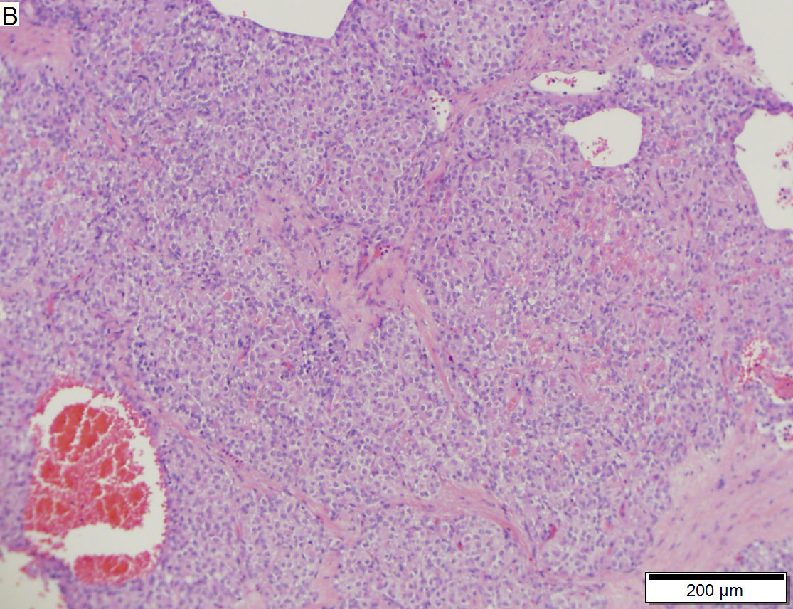
- Solid sheets of cells, focally dyscohesive.
- Eosinophilic cytoplasm.
- Occasionally clear cytoplasm.[4]
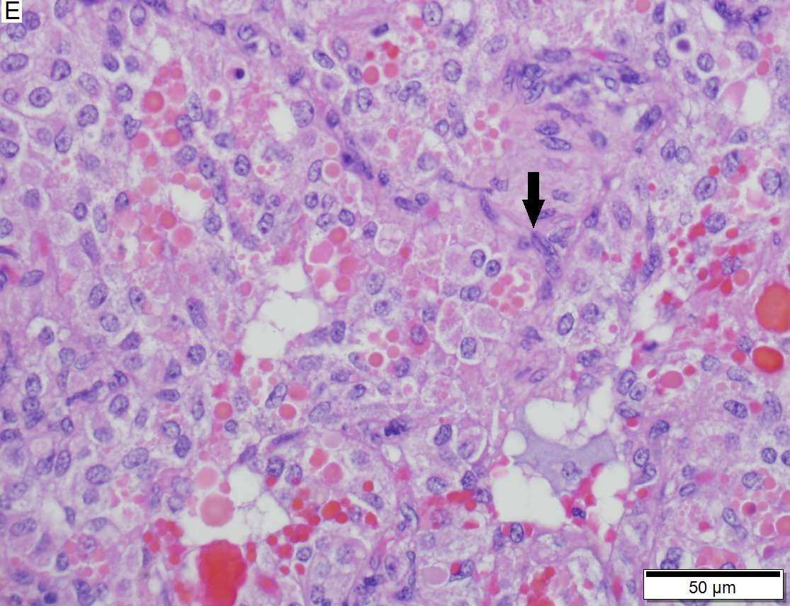
- Focal eosinophilic (intracytoplasmic) globules - key feature.
- Uniform nuclei with occasional nuclear grooves.
- +/-Necrosis - creating spaces/cavities.
- +/-Cholesterol clefts.[5]
DDx:
- Pancreatic pseudocyst.
- Cystadenoma.
- Cystadenocarcinoma - see invasive ductal carcinoma of the pancreas.
- Pancreatic neuroendocrine tumour - may have cytoplasmic vacuolation, hyaline globules.[4]
Images
www:
Solid-pseudopapillary neoplasm of pancreas. A. Tumor with prominent blood vessels and small cyst like spaces contrasts with normal pancreatic acini at right. B. Cohesive cell population. C. More dishesive cell population. D. Vascular pseudopapillae with hyalinized stroma. E. Aggregates of variably sized hyaline globules. Arrow points to longitudinal nuclear groove. F. Sometimes, as here, the cells show varied nuclear sizes and shapes. The cytoplasm, lightly eosinophilic to cleared, can have vacuoles (arrows).
IHC
Features:[4]
- Beta-catenin +ve ~100% (cytoplasmic & nuclear).
- E-cadherin +ve ~100% (cytoplasmic), -ve (membrane); antibody dependent.
- CD10 +ve ~ 80% (cytoplasmic + dot-like) key.
- Synaptophysin +ve (weak cytoplasmic) ~70%.
- Progesterone receptor +ve (nuclear) key.
Others:
- CD56 +ve.
- Chromogranin -ve.
Memory device PCB: PR (nuclear), CD10 (cytoplasmic), beta-catenin (cytoplasmic & nuclear).
See also
References
- ↑ URL: http://brighamrad.harvard.edu/Cases/bwh/hcache/360/full.html. Accessed on: 31 October 2011.
- ↑ Iacobuzio-Donahue, Christine A.; Montgomery, Elizabeth A. (2005). Gastrointestinal and Liver Pathology: A Volume in the Foundations in Diagnostic Pathology Series (1st ed.). Churchill Livingstone. pp. 493. ISBN 978-0443066573.
- ↑ Iacobuzio-Donahue, Christine A.; Montgomery, Elizabeth A. (2005). Gastrointestinal and Liver Pathology: A Volume in the Foundations in Diagnostic Pathology Series (1st ed.). Churchill Livingstone. pp. 493-5. ISBN 978-0443066573.
- ↑ 4.0 4.1 4.2 Serra S, Chetty R (November 2008). "Revision 2: an immunohistochemical approach and evaluation of solid pseudopapillary tumour of the pancreas". J. Clin. Pathol. 61 (11): 1153–9. doi:10.1136/jcp.2008.057828. PMID 18708424. http://jcp.bmj.com/content/61/11/1153.
- ↑ Abad Licham, M.; Sanchez Lihon, J.; Celis Zapata, J.. "[Pseudopapillary solid tumor of pancreas in the INEN].". Rev Gastroenterol Peru 28 (4): 356-61. PMID 19156179.


