Small bowel pseudomelanosis
(Redirected from Pseudomelanosis ileum)
Jump to navigation
Jump to search
| Small bowel pseudomelanosis | |
|---|---|
| Diagnosis in short | |
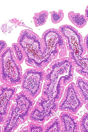
 Small bowel pseudomelanosis. H&E stain. | |
|
| |
| Synonyms | pseudomelanosis duodeni (for duodenum) |
|
| |
| LM | dark pigment in the lamina propria macrophages |
| Stains | iron stain +ve |
| Site | small bowel, stomach |
|
| |
| Associated Dx | iron deficiency anemia, end-stage renal disease, hypertension |
| Clinical history | iron supplementation |
| Prevalence | rare |
| Endoscopy | +/-dark spots |
| Prognosis | benign |
Small bowel pseudomelanosis is benign change of the small bowel that mimics melanin deposition.
In the duodenum it is known as pseudomelanosis duodeni.
Pseudomelanosis is described in the stomach, duodenum and jejunum.[1]
General
Associations in a series of 17 individuals:[4]
- Hypertension ~90% of cases.
- Iron supplementation ~75% of cases.
- End-stage renal disease ~60% of cases.
Note:
- The associations are different than for melanosis coli.
Gross/endoscopic
- Dark spots ~35% of cases.[4]
Microscopic
Features:
- Dark pigment in the lamina propria macrophages.
Images
Case 1
Case 2
www
Stains
- Prussian blue +ve ~80% of cases.[4]
Sign out
Small bowel
A. Stomach, Partial Excision as Part of a Sleeve Gastrectomy:
- Stomach wall within normal limits.

B. Small Bowel Wall, Partial Excision as Part of a Sleeve Gastrectomy:
- Small bowel wall with iron containing macrophages in the mucosa,
  known as "pseudomelanosis", see comment.
- NEGATIVE for active inflammation.
- NEGATIVE for dysplasia and NEGATIVE for malignancy.

Comment:
Pseudomelanosis may be seen in the context of iron supplementation, hypertension
or chronic renal failure.

The macrophages are positive with an iron stain.
Duodenum
Duodenum, Biopsy:
- Small bowel mucosa with pseudomelanosis, otherwise within
  normal limits, see comment.

Comment:
Pseudomelanosis of the duodenum (pseudomelanosis duodeni) may be seen in the context of iron supplementation, hypertension or chronic renal failure. Endoscopically, it may manifest as dark spots.

The pigmented cells are POSITIVE with an iron stain, NEGATIVE with a PAS stain and NEGATIVE with a Fontana-Masson stain; these findings are in keeping pseudomelanosis duodeni.
See also
References
- ↑ Rustagi, T.; Mansoor, MS.; Gibson, JA.; Kapadia, CR. (Feb 2015). "Pseudomelanosis of stomach, duodenum, and jejunum.". J Clin Gastroenterol 49 (2): 124-6. doi:10.1097/MCG.0000000000000081. PMID 24492404.
- ↑ de Magalhães Costa, MH.; Fernandes Pegado, Mda G.; Vargas, C.; Castro, ME.; Madi, K.; Nunes, T.; Zaltman, C. (Mar 2012). "Pseudomelanosis duodeni associated with chronic renal failure.". World J Gastroenterol 18 (12): 1414-6. doi:10.3748/wjg.v18.i12.1414. PMID 22493558.
- ↑ Lin, HJ.; Tsay, SH.; Chiang, H.; Tsai, YT.; Lee, SD.; Yeh, YS.; Lo, GH. (Apr 1988). "Pseudomelanosis duodeni. Case report and review of literature.". J Clin Gastroenterol 10 (2): 155-9. PMID 2458404.
- ↑ 4.0 4.1 4.2 Giusto, D.; Jakate, S. (Feb 2008). "Pseudomelanosis duodeni: associated with multiple clinical conditions and unpredictable iron stainability - a case series.". Endoscopy 40 (2): 165-7. doi:10.1055/s-2007-995472. PMID 18253910.


