File:Gallbladder cholesterolosis micro.jpg
Jump to navigation
Jump to search

Size of this preview: 800 × 600 pixels. Other resolution: 2,048 × 1,536 pixels.
Original file (2,048 × 1,536 pixels, file size: 924 KB, MIME type: image/jpeg)
Summary
| Description |
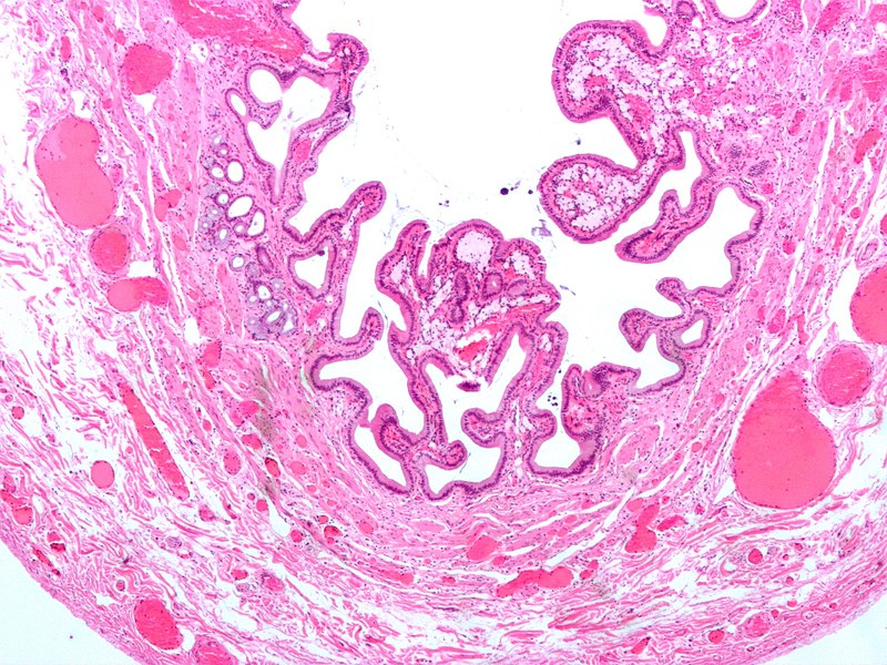
English: Micrograph of gallbladder with cholesterolosis. Cholecystectomy specimen. H&E stain. Low power magnification.
Dead lipid laden macrophages (foam cells) are seen in the finger-like projections into the gallbladder lumen. It should be apparent that this is gallbladder, as no muscularis mucosae is present (as elsewhere in the gastrointestinal tract). The blood vessels are congested and the subserosa edematous. |
||
| Date | |||
| Source | Own work | ||
| Author | Nephron | ||
| Permission (Reusing this file) |
I, the copyright holder of this work, hereby publish it under the following licenses: This file is licensed under the Creative Commons Attribution-Share Alike 3.0 Unported license.
You may select the license of your choice. |

File history
Click on a date/time to view the file as it appeared at that time.
| Date/Time | Thumbnail | Dimensions | User | Comment | |
|---|---|---|---|---|---|
| current | 08:21, 6 February 2009 | | 2,048 × 1,536 (924 KB) | Nephron | {{Information |Description={{en|1=Micrograph of '''gallbladder with cholesterolosis'''. Cholecystectomy specimen. H&E stain. Low power magnification. Dead lipid laden macrophages (foam cells) are seen in the finger-like projections into the gallbladder l |
File usage
The following 2 pages use this file: