Auer rod
(Redirected from Auer body)
Jump to navigation
Jump to search
.jpg)
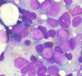
Micrograph showing an Auer rod in a blast. Wright stain. (WC)
Auer rod a microscopic finding typical of some types of acute myeloid leukemia and seen in myelodysplastic syndromes.
It may be referred to as an Auer body.
General
Microscopic
Features:
- Needle-like cytoplasmic bodies - classic appearance.[3]
- Other shapes: comma-like, diamond-like, rectangular, rarely glandular or corkscrew-like.
DDx:
- Auer-rod like inclusions - may be seen in multiple myeloma[4] or lymphoma cells.[2]
Images
Auer rods. (WC)
- Faggot cell in AML-M3.jpg
Abundant Auer rods in a Faggot cell. (WC)

See als
References
- ↑ Yoshida, Y.; Oguma, S.; Ohno, H.; Auer, J. (May 2009). "John Auer and Auer rods; controversies revisited.". Leuk Res 33 (5): 614-6. doi:10.1016/j.leukres.2008.09.014. PMID 18947869.
- ↑ 2.0 2.1 Groom, DA.; Wong, D.; Brynes, RK.; Macaulay, LK. (Jul 1991). "Auer rod-like inclusions in circulating lymphoma cells.". Am J Clin Pathol 96 (1): 111-5. PMID 1712539.
- ↑ ACKERMAN, GA. (Sep 1950). "Microscopic and histochemical studies on the Auer bodies in leukemic cells.". Blood 5 (9): 847-63. PMID 15434012.
- ↑ Abdulsalam, AH.; Bain, BJ. (Mar 2014). "Auer-rod like inclusions in multiple myeloma.". Am J Hematol 89 (3): 338. doi:10.1002/ajh.23648. PMID 24338920.