Alpha-1 antitrypsin deficiency
| Alpha-1 antitrypsin deficiency | |
|---|---|
| Diagnosis in short | |
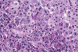 Alpha-1 AT deficiency. PASD stain. | |
|
| |
| Synonyms | alpha1-antiprotease inhibitor deficiency |
|
| |
| LM | +/-pink globules in zone 1 (periportal), +/-fibrosis or cirrhosis |
| Stains | PASD +ve (pink globules in zone 1) - not seen in children |
| IHC | A1-AT +ve globules |
| Site | liver - see medical liver disease, lung - see emphysema |
|
| |
| Prevalence | uncommon (1 in 2000-5000) |
| Radiology | emphysematous changes (chest x-ray) |
Alpha-1 antitrypsin deficiency, abbreviated A1-AT, is a relatively common genetic condition that causes lung and liver pathology.
It is also known as alpha1-antiprotease inhibitor deficiency.
This article deals with the liver pathology. The lung pathology is panlobular emphysema and covered in the emphysema article.
General
Etiology:
- Genetic defect.
- Prevalence 1 in 2000-5000.[1]
Causes:
- Lung and liver injury.
- Lung -> panlobular emphysema.
Microscopic
Features:
- Pink globules in zone 1 (periportal).
- Globules not seen in children.
- May not be present in late stage (cirrhotic).
- Best seen on PASD stain.
- Can be seen on H&E -- if one looks carefully.
Note:
- The pink globules may be seen in the context of cirrhosis; cases should be confirmed with IHC.
Images
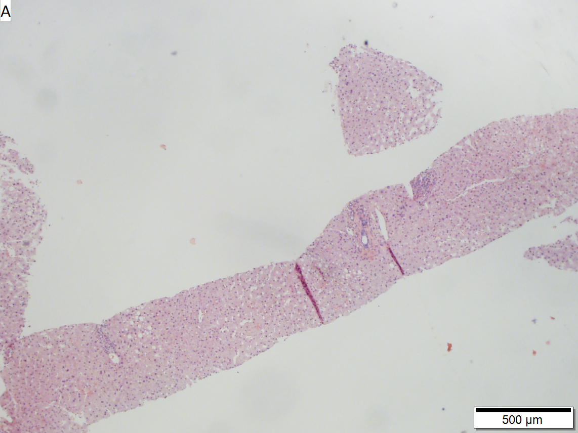

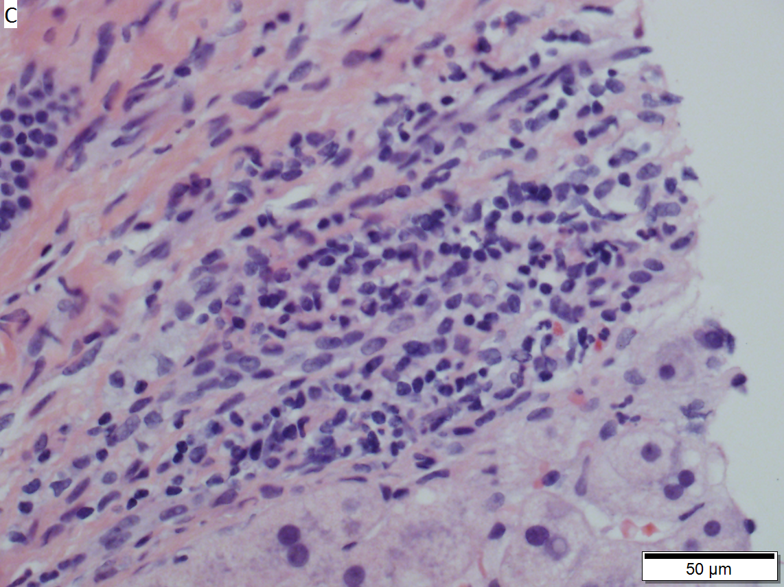

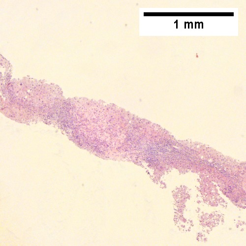
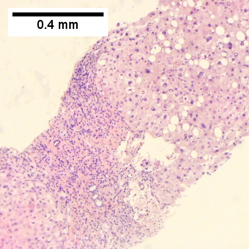
Alpha 1 anti-trypsin deficiency in 29 year old woman. A. At low power, there is focal steatosis and mild inflammation of portal triads. B. Foci of steatosis show rare tufts of ballooning degeneration (arrow). C. A few triads had infiltrates of macrophages and lymphocytes, none with definite interface hepatitis; note the space between the hepatocytes and the inflammatory cells. D. PAS-D showed splotches of hepatocytes with PAS-D granules. The patient was already known to have A1AT deficiency.
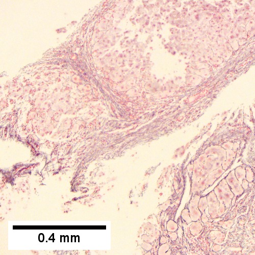
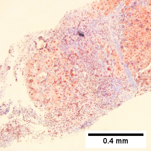
Alpha 1 Antitrypsin deficiency with cirrhosis. A. Dark inflamed bands bound hepatocyte nodules. B. Steatosis afflicts some hepatocyte areas, within inflamed band lie proliferated bile ductules. C. Reticulin shows regenerative (more than 2 nuclei per cord) nodules (black lines without definite directionality) amid bands. D. Trichrome shows scar. E. Hepatocytes show steatosis, foamy degeneration, and intranuclear inclusions. F. PAS-D shows A1AT cytoplasmic granules; in non-A1AT cirrhosis, these granules would neither be in so many hepatocytes nor, in general, as large.
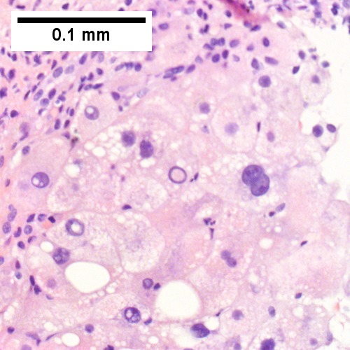
Alpha 1 anti-trypsin (A1AT) granules in cirrhosis, not due to A1AT deficiency; A1AT level was normal.
Alpha 1 anti-trypsin (A1AT) granules in cirrhosis, not due to A1AT deficiency; A1AT level was normal.
Alpha 1 anti-trypsin (A1AT) granules in cirrhosis, not due to A1AT deficiency; A1AT level was normal.
Alpha 1 anti-trypsin (A1AT) granules in cirrhosis, not due to A1AT deficiency; A1AT level was normal.
Alpha 1 anti-trypsin (A1AT) granules in cirrhosis, not due to A1AT deficiency; A1AT level was normal.
Alpha 1 anti-trypsin (A1AT) granules in cirrhosis, not due to A1AT deficiency; A1AT level was normal.
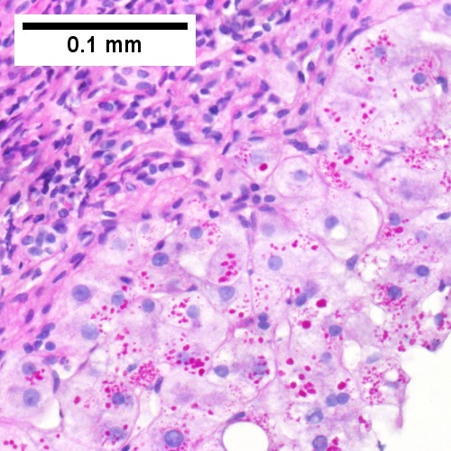
Alpha 1 anti-trypsin (A1AT) granules in cirrhosis, not due to A1AT deficiency; A1AT level was normal. A. Low power shows an inflamed foci, bands, and hepatocyte regions. B. Trichrome confirms blue fibrosis about hepatocyte isles. C. Proliferated bile ductules (black arrows) border hepatocytes with glycogenated nuclei (green arrows), consistent with the history of diabetes. D. Reticulin shows two-cell thick regenerated cords (green arrows) and circles for rosettes (black arrows). E. PAS with diastase stain shows occasional small red granules (arrows) and red dust in cytoplasm of hepatocytes near edge of regenerative nodule. E. A1AT immunostain shows occasional small brown granules (arrows) in cytoplasm of hepatocytes near edge of regenerative nodule.
www:
Stains
IHC
- A1-AT +ve globules.[3]
See also
References
- ↑ Stoller, JK.; Aboussouan, LS. (Sep 2011). "A Review of Alpha-1 Antitrypsin Deficiency.". Am J Respir Crit Care Med. doi:10.1164/rccm.201108-1428CI. PMID 21960536.
- ↑ 2.0 2.1 Qizilbash, A.; Young-Pong, O. (Jun 1983). "Alpha 1 antitrypsin liver disease differential diagnosis of PAS-positive, diastase-resistant globules in liver cells.". Am J Clin Pathol 79 (6): 697-702. PMID 6189389.
- ↑ Theaker, JM.; Fleming, KA. (Jan 1986). "Alpha-1-antitrypsin and the liver: a routine immunohistological screen.". J Clin Pathol 39 (1): 58-62. PMID 3512609.

