Difference between revisions of "Walthard cell rest"
Jump to navigation
Jump to search
m (fix redirect) |
m (→General: fix another typo) |
||
| (15 intermediate revisions by the same user not shown) | |||
| Line 1: | Line 1: | ||
{{ Infobox diagnosis | |||
| Name = {{PAGENAME}} | |||
| Image = Walthard cell rest - very low mag.jpg | |||
| Width = | |||
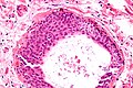
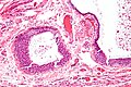
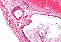
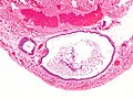
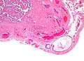
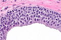
| Caption = A cystic Walthard cell rest of the [[fallopian tube]]. [[H&E stain]]. | |||
| Synonyms = | |||
| Micro = collection of eosinophilic (i.e. pink) cuboidal cells - usually solid, may be cystic; elliptical nucleus with single groove along major axis - "coffee bean" nucleus | |||
| Subtypes = | |||
| LMDDx = [[Brenner tumour]] | |||
| Stains = | |||
| IHC = | |||
| EM = | |||
| Molecular = | |||
| IF = | |||
| Gross = | |||
| Grossing = | |||
| Site = [[Fallopian tube]], [[testis]] | |||
| Assdx = | |||
| Syndromes = none | |||
| Clinicalhx = | |||
| Signs = none | |||
| Symptoms = | |||
| Prevalence = common | |||
| Bloodwork = | |||
| Rads = | |||
| Endoscopy = | |||
| Prognosis = benign | |||
| Other = | |||
| ClinDDx = | |||
| Tx = none | |||
}} | |||
'''Walthard cell rest''', also '''Walthard cell nest''', is a benign finding often seen in [[gynecologic pathology]]. | |||
==General== | |||
*Benign. | |||
*Typically an incidental finding. | |||
*May be found in the [[testis]].<ref name=pmid15502808>{{cite journal |author=Amin MB |title=Selected other problematic testicular and paratesticular lesions: rete testis neoplasms and pseudotumors, mesothelial lesions and secondary tumors |journal=Mod. Pathol. |volume=18 Suppl 2 |issue= |pages=S131–45 |year=2005 |month=February |pmid=15502808 |doi=10.1038/modpathol.3800314 |url=}}</ref> | |||
===Epidemiology=== | |||
*Thought to be related to [[Brenner tumour]]. | |||
==Microscopic== | |||
Features:<ref name=Ref_GP332>{{Ref_GP|332}}</ref> | |||
*Collection of eosinophilic (i.e. pink) cuboidal cells; usually solid, may be cystic. | |||
*Elliptical nucleus with single groove along major axis; [[coffee bean nucleus|"coffee bean" nucleus]] -- '''key feature'''. | |||
Location: | |||
*Usually in soft tissue of the uterine tube. | |||
DDx: | |||
*[[Brenner tumour]] - consist of (multiple) nests with a surrounding fibromatous stroma.<ref name=pmid25281026>{{Cite journal | last1 = Roma | first1 = AA. | last2 = Masand | first2 = RP. | title = Ovarian Brenner tumors and Walthard nests: a histologic and immunohistochemical study. | journal = Hum Pathol | volume = 45 | issue = 12 | pages = 2417-22 | month = Dec | year = 2014 | doi = 10.1016/j.humpath.2014.08.003 | PMID = 25281026 }}</ref> | |||
===Images=== | |||
====Fallopian tube==== | |||
<gallery> | |||
Image:Walthard cell rest - very high mag.jpg | WCR - very high mag. | |||
Image:Walthard cell rest - high mag.jpg | WCR - high mag. | |||
Image:Walthard cell rest - intermed mag.jpg | WCR - intermed. mag. | |||
Image:Walthard cell rest - low mag.jpg | WCR - low mag. | |||
Image:Walthard cell rest - very low mag.jpg | WCR - very low mag. | |||
</gallery> | |||
====Testis==== | |||
<gallery> | |||
Image:Walthard cell rest testis -- low mag.jpg | Low mag. | |||
Image:Walthard cell rest testis -- intermed mag.jpg | Intermed. mag. | |||
Image:Walthard cell rest testis -- high mag.jpg | High mag. | |||
Image:Walthard cell rest testis -- very high mag.jpg | Very high mag. | |||
</gallery> | |||
==See also== | |||
*[[Gynecologic pathology]]. | |||
*[[Fallopian tube]]. | |||
==References== | |||
{{Reflist|1}} | |||
[[Category:Gynecologic pathology]] | |||
Latest revision as of 19:23, 7 March 2016
| Walthard cell rest | |
|---|---|
| Diagnosis in short | |
 A cystic Walthard cell rest of the fallopian tube. H&E stain. | |
|
| |
| LM | collection of eosinophilic (i.e. pink) cuboidal cells - usually solid, may be cystic; elliptical nucleus with single groove along major axis - "coffee bean" nucleus |
| LM DDx | Brenner tumour |
| Site | Fallopian tube, testis |
|
| |
| Syndromes | none |
|
| |
| Signs | none |
| Prevalence | common |
| Prognosis | benign |
| Treatment | none |
Walthard cell rest, also Walthard cell nest, is a benign finding often seen in gynecologic pathology.
General
Epidemiology
- Thought to be related to Brenner tumour.
Microscopic
Features:[2]
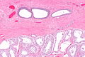
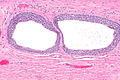
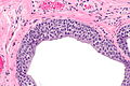
- Collection of eosinophilic (i.e. pink) cuboidal cells; usually solid, may be cystic.
- Elliptical nucleus with single groove along major axis; "coffee bean" nucleus -- key feature.
Location:
- Usually in soft tissue of the uterine tube.
DDx:
- Brenner tumour - consist of (multiple) nests with a surrounding fibromatous stroma.[3]
Images
Fallopian tube
WCR - very high mag.
WCR - high mag.
WCR - intermed. mag.
WCR - low mag.
WCR - very low mag.


Testis
Low mag.
Intermed. mag.
High mag.
Very high mag.


See also
References
- ↑ Amin MB (February 2005). "Selected other problematic testicular and paratesticular lesions: rete testis neoplasms and pseudotumors, mesothelial lesions and secondary tumors". Mod. Pathol. 18 Suppl 2: S131–45. doi:10.1038/modpathol.3800314. PMID 15502808.
- ↑ Nucci, Marisa R.; Oliva, Esther (2009). Gynecologic Pathology: A Volume in Foundations in Diagnostic Pathology Series (1st ed.). Churchill Livingstone. pp. 332. ISBN 978-0443069208.
- ↑ Roma, AA.; Masand, RP. (Dec 2014). "Ovarian Brenner tumors and Walthard nests: a histologic and immunohistochemical study.". Hum Pathol 45 (12): 2417-22. doi:10.1016/j.humpath.2014.08.003. PMID 25281026.