Difference between revisions of "Walthard cell rest"
Jump to navigation
Jump to search
(→Images) |
|||
| Line 49: | Line 49: | ||
===Images=== | ===Images=== | ||
====Fallopian tube==== | |||
<gallery> | <gallery> | ||
Image:Walthard cell rest - very high mag.jpg | WCR - very high mag. | Image:Walthard cell rest - very high mag.jpg | WCR - very high mag. | ||
| Line 55: | Line 56: | ||
Image:Walthard cell rest - low mag.jpg | WCR - low mag. | Image:Walthard cell rest - low mag.jpg | WCR - low mag. | ||
Image:Walthard cell rest - very low mag.jpg | WCR - very low mag. | Image:Walthard cell rest - very low mag.jpg | WCR - very low mag. | ||
</gallery> | |||
====Testis==== | |||
<gallery> | |||
Image:Walthard cell rest testis -- low mag.jpg | Low mag. | |||
Image:Walthard cell rest testis -- intermed mag.jpg | Intermed. mag. | |||
Image:Walthard cell rest testis -- high mag.jpg | High mag. | |||
Image:Walthard cell rest testis -- very high mag.jpg | Very high mag. | |||
</gallery> | </gallery> | ||
Revision as of 04:46, 30 July 2014
| Walthard cell rest | |
|---|---|
| Diagnosis in short | |
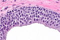
 Walthard cell rest. H&E stain. | |
|
| |
| LM | collection of eosinophilic (i.e. pink) cuboidal cells - usually solid, may be cystic; elliptical nucleus with single groove along major axis - "coffee bean" nucleus |
| Site | Fallopian tube, testis |
|
| |
| Syndromes | none |
|
| |
| Signs | none |
| Prevalence | common |
| Prognosis | benign |
| Treatment | none |
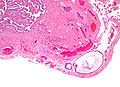
Walthard cell rest, also Walthard cell nest, is a benign finding in often seen in gynecologic pathology.
General
Epidemiology
- Thought to be related to Brenner tumour.
Microscopic
Features:[2]
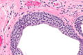
- Collection of eosinophilic (i.e. pink) cuboidal cells; usually solid, may be cystic.
- Elliptical nucleus with single groove along major axis; "coffee bean" nucleus -- key feature.
Location:
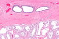
- Usually in soft tissue of the uterine tube.
Images
Fallopian tube
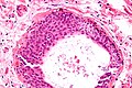
WCR - very high mag.
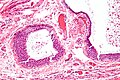
WCR - high mag.
WCR - intermed. mag.
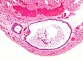
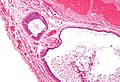
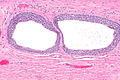
WCR - low mag.
WCR - very low mag.


Testis
Low mag.
Intermed. mag.
High mag.
Very high mag.


See also
References
- ↑ Amin MB (February 2005). "Selected other problematic testicular and paratesticular lesions: rete testis neoplasms and pseudotumors, mesothelial lesions and secondary tumors". Mod. Pathol. 18 Suppl 2: S131–45. doi:10.1038/modpathol.3800314. PMID 15502808.
- ↑ Nucci, Marisa R.; Oliva, Esther (2009). Gynecologic Pathology: A Volume in Foundations in Diagnostic Pathology Series (1st ed.). Churchill Livingstone. pp. 332. ISBN 978-0443069208.