Difference between revisions of "Verrucous carcinoma"
Jump to navigation
Jump to search
(split-out) Tag: Removed redirect |
|||
| (One intermediate revision by the same user not shown) | |||
| Line 19: | Line 19: | ||
===Images=== | ===Images=== | ||
<gallery> | |||
Image: Micrograph of penile verrucous carcinoma - 20x.jpg | Penile VC - Low Mag. (WC) | |||
Image: Micrograph of penile verrucous carcinoma - 200x.jpg | Penile VC - High Mag. (WC) | |||
</gallery> | |||
*[http://www.juniordentist.com/wp-content/uploads/2011/06/Verrucous-carcinoma-histology.jpg Verrucous carcinoma (juniordentist.com)].<ref>URL: [http://www.juniordentist.com/verrucous-carcinoma.html http://www.juniordentist.com/verrucous-carcinoma.html]. Accessed on: 3 April 2012.</ref> | *[http://www.juniordentist.com/wp-content/uploads/2011/06/Verrucous-carcinoma-histology.jpg Verrucous carcinoma (juniordentist.com)].<ref>URL: [http://www.juniordentist.com/verrucous-carcinoma.html http://www.juniordentist.com/verrucous-carcinoma.html]. Accessed on: 3 April 2012.</ref> | ||
*[http://www.pathologyoutlines.com/images/prostate/psverrucous_scc_01.jpg Verrucous SCC (pathologyoutlines.com)]. | *[http://www.pathologyoutlines.com/images/prostate/psverrucous_scc_01.jpg Verrucous SCC (pathologyoutlines.com)]. | ||
| Line 33: | Line 38: | ||
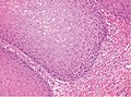
====Micro==== | ====Micro==== | ||
The sections show superficial squamous mucosa with keratinization and church spire-like structures. Mild basal cellular changes are present. Significant nuclear atypia is absent. Minimal stroma with mild inflammation is present. | The sections show superficial squamous mucosa with keratinization and church spire-like structures. Mild basal cellular changes are present. Significant epithelial nuclear atypia is absent. Minimal stroma with mild inflammation is present. | ||
==See also== | ==See also== | ||
Latest revision as of 14:32, 19 March 2024
Verrucous carcinoma, also verrucous squamous cell carcinoma, is an uncommon variant of squamous cell carcinoma that may be challenging to diagnose.
General
- Good prognosis.
- Histomorphologically deceptively bland, i.e. non-malignant appearing.
- Wart-like (verruca = wart).
- The clinical history, e.g. huge verrucous cancer, is often important for making the diagnosis.
Microscopic
Features:
- Exophytic growth.
- Well-differentiated.
- "Glassy" appearance.
- Pushing border - described "elephant feet".
DDx:
Images

Penile VC - Low Mag. (WC)
Penile VC - High Mag. (WC)


Sign out
Biopsy
Mouth Lesion, Hard Palate, Biopsy:
	- Squamous mucosa with keratinization and verrucous features, see comment.

Comment:
A clinic note describes a 2 cm mass of the hard palate. The clinical impression is noted. Correlation with imaging and the clinical features is recommended.
Micro
The sections show superficial squamous mucosa with keratinization and church spire-like structures. Mild basal cellular changes are present. Significant epithelial nuclear atypia is absent. Minimal stroma with mild inflammation is present.
See also
References
- ↑ URL: http://www.juniordentist.com/verrucous-carcinoma.html. Accessed on: 3 April 2012.