Difference between revisions of "Verruca vulgaris"
(redirect) |
|||
| (3 intermediate revisions by the same user not shown) | |||
| Line 1: | Line 1: | ||
{{ Infobox diagnosis | |||
| Name = {{PAGENAME}} | |||
| Image = Verruca_vulgaris_-_very_low_mag.jpg | |||
| Width = | |||
| Caption = Verruca vulgaris. [[H&E stain]]. | |||
| Micro = papillomatous hyperplasia (rete ridges long & curve inward), hyperkeratosis, hypergranulosis, large blood vessels at the dermal-epidermal junction, +/-viral changes - perinuclear halo, nucleus small and hyperchromatic (virtually diagnostic when present), +/-binucleation | |||
| Subtypes = | |||
| LMDDx = [[Squamous cell carcinoma of the skin|Squamous cell carcinoma]], hypertrophic [[actinic keratosis]], actinic keratosis with superimposed [[lichen simplex chronicus]], [[seborrheic keratosis]], [[condyloma acuminatum]] | |||
| Stains = | |||
| IHC = | |||
| EM = | |||
| Molecular = | |||
| IF = | |||
| Gross = | |||
| Grossing = | |||
| Site = [[skin]] - classically hands | |||
| Assdx = | |||
| Syndromes = | |||
| Clinicalhx = | |||
| Signs = | |||
| Symptoms = | |||
| Prevalence = very common | |||
| Bloodwork = | |||
| Rads = | |||
| Endoscopy = | |||
| Prognosis = benign | |||
| Other = | |||
| ClinDDx = | |||
}} | |||
'''Verruca vulgaris''', also known as '''common wart''', is a common [[non-malignant skin disease]] caused by certain subtypes of the [[human papilloma virus]]. | |||
==General== | |||
*Etiology - [[HPV]]. | |||
*Very common. | |||
Notes: | |||
*Related to [[condyloma acuminatum]]. | |||
==Gross== | |||
Features: | |||
*[[Papule]] or [[plaque]] with granular surface. | |||
*Classic location: hand. | |||
Images: | |||
*[http://commons.wikimedia.org/wiki/File:Porea_doet_djin_wi.jpg Wart (WC)]. | |||
*[http://commons.wikimedia.org/wiki/File:Wart_ASA_animated.gif Wart - animated (WC)]. | |||
==Microscopic== | |||
Features:<ref>URL: [http://missinglink.ucsf.edu/lm/DermatologyGlossary/verruca_vulgaris.html http://missinglink.ucsf.edu/lm/DermatologyGlossary/verruca_vulgaris.html]. Accessed on: 14 July 2010.</ref> | |||
*Hyperkeratosis (more keratin - thick stratum corneum) - in "columns"; keratin in separate towers - ''not'' a flat thick sheet. | |||
*Hypergranulosis (thicker stratum granulosum). | |||
*''Papillomatous hyperplasia'': | |||
**Rete ridge lengthening (~7-10x normal) and thickening. | |||
**Rete ridge curvature toward the centre of the lesion (like the roads to the ''Palace of Versailles'') - '''important'''. | |||
*Large blood vessels at the dermal-epidermal junction - between the rete ridges. | |||
*+/-Viral changes - perinuclear halo, nucleus small and hyperchromatic<ref name=Ref_Derm106-7>{{Ref Derm|106-7}}</ref> - virtually '''diagnostic''' when present. | |||
**+/-Binucleation. | |||
Memory device: there is more of everything - more s. corneum, s. granulosum, s. spinosum, longer rete ridges, more (larger) blood vessels. | |||
DDx: | |||
*[[Squamous cell carcinoma of the skin|Squamous cell carcinoma]] - nuclear atypia full thickness and often more pronounced. | |||
*Hypertrophic [[actinic keratosis]] - often accompanied by solar elastosis. | |||
**Actinic keratosis with superimposed [[lichen simplex chronicus]].<ref name=Ref_Derm353>{{Ref Derm|353}}</ref> | |||
*[[Seborrheic keratosis]] - may have focal clear cell changes. | |||
*[[Condyloma acuminatum]] - genital region. | |||
===Images=== | |||
<gallery> | |||
Image:Verruca_vulgaris_-_very_low_mag.jpg | Verruca vulgaris - very low mag. (WC) | |||
Image:Verruca_vulgaris_-_intermed_mag.jpg | Verruca vulgaris - intermed mag. (WC) | |||
</gallery> | |||
www: | |||
*[http://alf3.urz.unibas.ch/pathopic/getpic-fra.cfm?id=4528 Verruca vulgaris (unibas.ch)]. | |||
==Sign out== | |||
<pre> | |||
SKIN LESION, RIGHT LOWER LEG, SHAVE BIOPSY: | |||
- VERRUCA VULGARIS. | |||
</pre> | |||
===Micro=== | |||
The sections show skin with elongated rete ridges, acanthosis, hypergranulosis, hyperkeratosis in vertical columns, focal parakeratosis, dilated blood vessels at the dermal-epidermal junction and koilocytic change. Mild basilar nuclear atypia is present. | |||
====Without koilocytes==== | |||
The sections show skin with elongated rete ridges that curve toward the centre of the lesion, acanthosis, hypergranulosis, hyperkeratosis in vertical columns, and dilated blood vessels at the dermal-epidermal | |||
junction. Minimal basilar nuclear enlargement is present. No definite koilocytic change is | |||
apparent. | |||
No parakeratosis is identified. Mild solar elastosis is identified. No melanocytic nests | |||
are apparent. Mitotic activity is not apparent. | |||
==See also== | |||
*[[Non-malignant skin disease]]. | |||
==References== | |||
{{Reflist|2}} | |||
[[Category:Diagnosis]] | |||
[[Category:Non-malignant skin disease]] | |||
Latest revision as of 12:06, 11 October 2013
| Verruca vulgaris | |
|---|---|
| Diagnosis in short | |
 Verruca vulgaris. H&E stain. | |
|
| |
| LM | papillomatous hyperplasia (rete ridges long & curve inward), hyperkeratosis, hypergranulosis, large blood vessels at the dermal-epidermal junction, +/-viral changes - perinuclear halo, nucleus small and hyperchromatic (virtually diagnostic when present), +/-binucleation |
| LM DDx | Squamous cell carcinoma, hypertrophic actinic keratosis, actinic keratosis with superimposed lichen simplex chronicus, seborrheic keratosis, condyloma acuminatum |
| Site | skin - classically hands |
|
| |
| Prevalence | very common |
| Prognosis | benign |
Verruca vulgaris, also known as common wart, is a common non-malignant skin disease caused by certain subtypes of the human papilloma virus.
General
- Etiology - HPV.
- Very common.
Notes:
- Related to condyloma acuminatum.
Gross
Features:
Images:
Microscopic
Features:[1]
- Hyperkeratosis (more keratin - thick stratum corneum) - in "columns"; keratin in separate towers - not a flat thick sheet.
- Hypergranulosis (thicker stratum granulosum).
- Papillomatous hyperplasia:
- Rete ridge lengthening (~7-10x normal) and thickening.
- Rete ridge curvature toward the centre of the lesion (like the roads to the Palace of Versailles) - important.
- Large blood vessels at the dermal-epidermal junction - between the rete ridges.
- +/-Viral changes - perinuclear halo, nucleus small and hyperchromatic[2] - virtually diagnostic when present.
- +/-Binucleation.
Memory device: there is more of everything - more s. corneum, s. granulosum, s. spinosum, longer rete ridges, more (larger) blood vessels.
DDx:
- Squamous cell carcinoma - nuclear atypia full thickness and often more pronounced.
- Hypertrophic actinic keratosis - often accompanied by solar elastosis.
- Actinic keratosis with superimposed lichen simplex chronicus.[3]
- Seborrheic keratosis - may have focal clear cell changes.
- Condyloma acuminatum - genital region.
Images
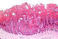
Verruca vulgaris - very low mag. (WC)
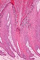
Verruca vulgaris - intermed mag. (WC)

www:
Sign out
SKIN LESION, RIGHT LOWER LEG, SHAVE BIOPSY:
- VERRUCA VULGARIS.
Micro
The sections show skin with elongated rete ridges, acanthosis, hypergranulosis, hyperkeratosis in vertical columns, focal parakeratosis, dilated blood vessels at the dermal-epidermal junction and koilocytic change. Mild basilar nuclear atypia is present.
Without koilocytes
The sections show skin with elongated rete ridges that curve toward the centre of the lesion, acanthosis, hypergranulosis, hyperkeratosis in vertical columns, and dilated blood vessels at the dermal-epidermal junction. Minimal basilar nuclear enlargement is present. No definite koilocytic change is apparent.
No parakeratosis is identified. Mild solar elastosis is identified. No melanocytic nests are apparent. Mitotic activity is not apparent.
See also
References
- ↑ URL: http://missinglink.ucsf.edu/lm/DermatologyGlossary/verruca_vulgaris.html. Accessed on: 14 July 2010.
- ↑ Busam, Klaus J. (2009). Dermatopathology: A Volume in the Foundations in Diagnostic Pathology Series (1st ed.). Saunders. pp. 106-7. ISBN 978-0443066542.
- ↑ Busam, Klaus J. (2009). Dermatopathology: A Volume in the Foundations in Diagnostic Pathology Series (1st ed.). Saunders. pp. 353. ISBN 978-0443066542.