Difference between revisions of "Urothelial papilloma"
Jump to navigation
Jump to search
(split out) |
|||
| (5 intermediate revisions by the same user not shown) | |||
| Line 1: | Line 1: | ||
{{ Infobox diagnosis | |||
| Name = {{PAGENAME}} | |||
| Image = Urothelial papilloma - alt -- intermed mag.jpg | |||
| Width = | |||
| Caption = Urothelial papilloma. [[H&E stain]]. | |||
| Synonyms = | |||
| Micro = papillary fronds with minimal branching or fusion, cytology and thickness of normal urothelium, no mitoses | |||
| Subtypes = | |||
| LMDDx = [[low grade papillary urothelial carcinoma]], [[PUNLMP]] | |||
| Stains = | |||
| IHC = | |||
| EM = | |||
| Molecular = | |||
| IF = | |||
| Gross = | |||
| Grossing = | |||
| Site = [[urothelium]] - typically [[urinary bladder]] | |||
| Assdx = | |||
| Syndromes = | |||
| Clinicalhx = | |||
| Signs = | |||
| Symptoms = | |||
| Prevalence = uncommon | |||
| Bloodwork = | |||
| Rads = | |||
| Endoscopy = | |||
| Prognosis = benign | |||
| Other = | |||
| ClinDDx = other papillary lesions | |||
| Tx = | |||
}} | |||
'''Urothelial papilloma''' is a rare benign lesion of the [[urothelium]]. | '''Urothelial papilloma''' is a rare benign lesion of the [[urothelium]]. | ||
| Line 8: | Line 39: | ||
*If the person has a history of a low grade papillary urothelial carcinoma... it is likely a low grade papillary urothelial carcinoma. | *If the person has a history of a low grade papillary urothelial carcinoma... it is likely a low grade papillary urothelial carcinoma. | ||
*These cases are a consensus diagnosis, i.e. you show it to a colleague... if they agree you can call it. | *These cases are a consensus diagnosis, i.e. you show it to a colleague... if they agree you can call it. | ||
==Gross== | |||
*Exophytic lesion. | |||
==Microscopic== | ==Microscopic== | ||
| Line 21: | Line 55: | ||
*[[Low grade papillary urothelial carcinoma]]. | *[[Low grade papillary urothelial carcinoma]]. | ||
*[[PUNLMP]]. | *[[PUNLMP]]. | ||
===Images=== | |||
<gallery> | |||
Image: Urothelial papilloma -- very low mag.jpg | UP - very low mag. | |||
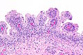
Image: Urothelial papilloma -- low mag.jpg | UP - low mag. | |||
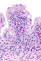
Image: Urothelial papilloma -- intermed mag.jpg | UP - intermed mag. | |||
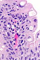
Image: Urothelial papilloma -- high mag.jpg | UP - high mag. | |||
Image: Urothelial papilloma -- very high mag.jpg | UP - very high mag. | |||
Image: Urothelial papilloma - alt 1 -- high mag.jpg | UP - high mag. | |||
Image: Urothelial papilloma - alt 2 -- high mag.jpg | UP - high mag. | |||
</gallery> | |||
==Sign out== | |||
<pre> | |||
Urinary Bladder, Biopsy: | |||
- Urothelial papilloma. | |||
</pre> | |||
===Microscopic=== | |||
The sections show superficial fragments of a papillary urothelial lesion. The cytology and thickness is that of normal urothelium. No proliferation is apparent. | |||
==See also== | ==See also== | ||
*[[Urothelium]]. | *[[Urothelium]]. | ||
*[[PUNLMP]]. | *[[PUNLMP]]. | ||
*[[Inverted urothelial papilloma]]. | |||
==References== | ==References== | ||
Latest revision as of 18:19, 19 September 2022
| Urothelial papilloma | |
|---|---|
| Diagnosis in short | |
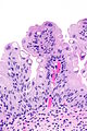
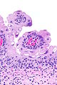
 Urothelial papilloma. H&E stain. | |
|
| |
| LM | papillary fronds with minimal branching or fusion, cytology and thickness of normal urothelium, no mitoses |
| LM DDx | low grade papillary urothelial carcinoma, PUNLMP |
| Site | urothelium - typically urinary bladder |
|
| |
| Prevalence | uncommon |
| Prognosis | benign |
| Clin. DDx | other papillary lesions |
Urothelial papilloma is a rare benign lesion of the urothelium.
General
Notes:
- If the person has a history of a low grade papillary urothelial carcinoma... it is likely a low grade papillary urothelial carcinoma.
- These cases are a consensus diagnosis, i.e. you show it to a colleague... if they agree you can call it.
Gross
- Exophytic lesion.
Microscopic
Features:[2]
- Papillary fronds.
- Minimal branching or fusion.
- Cytological features of normal urothelium.
- Normal urothelium approx. 2x the size of stromal lymphocytes.[3]
- No mitoses.
- Thickness < 7 cells.[citation needed]
DDx:
Images
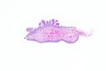
UP - very low mag.

UP - low mag.
UP - intermed mag.
UP - high mag.
UP - very high mag.
UP - high mag.
UP - high mag.


Sign out
Urinary Bladder, Biopsy:
	- Urothelial papilloma.
Microscopic
The sections show superficial fragments of a papillary urothelial lesion. The cytology and thickness is that of normal urothelium. No proliferation is apparent.
See also
References
- ↑ 1.0 1.1 Al Bashir, S.; Yilmaz, A.; Gotto, G.; Trpkov, K. (Jan 2014). "Long term outcome of primary urothelial papilloma: a single institution cohort.". Pathology 46 (1): 37-40. doi:10.1097/PAT.0000000000000029. PMID 24300727.
- ↑ Humphrey, Peter A; Dehner, Louis P; Pfeifer, John D (2008). The Washington Manual of Surgical Pathology (1st ed.). Lippincott Williams & Wilkins. pp. 310. ISBN 978-0781765275.
- ↑ Zhou, Ming; Magi-Galluzzi, Cristina (2006). Genitourinary Pathology: A Volume in Foundations in Diagnostic Pathology Series (1st ed.). Churchill Livingstone. pp. 161. ISBN 978-0443066771.