Urinary bladder amyloidosis
Jump to navigation
Jump to search
The printable version is no longer supported and may have rendering errors. Please update your browser bookmarks and please use the default browser print function instead.
| Urinary bladder amyloidosis | |
|---|---|
| Diagnosis in short | |
 Urinary bladder amyloidosis. H&E stain. | |
|
| |
| Synonyms | amyloidosis of the urinary bladder |
|
| |
| LM | lamina propria with amyloid (amorphous, paucicellular, pink material on H&E - classically "cracked") |
| LM DDx | fibrosis, fibrin, leiomyoma |
| Stains | Congo red +ve |
| Site | urinary bladder |
|
| |
| Prevalence | very rare |
| Radiology | thickened bladder wall |
| Clin. DDx | other urinary bladder tumours - esp. malignant ones |
Urinary bladder amyloidosis, more formally primary urinary bladder amyloidosis, is a rare benign condition of urinary bladder that can mimic cancer of the urinary bladder.[1]
General
- Primary bladder amyloidosis is rare - approximately 200 reported cases as of 2014.[1][2]
- Systemic amyloidosis should be excluded.[2]
- May clinically mimic bladder cancer.[1]
Gross
- Urinary bladder wall thickening.[1]
Microscopic
Features:
- Lamina propria with amyloid.
- Amyloid = amorphous, paucicellular material that is pink on H&E, classically has "cracked" appearance.
- "Cracked": irregular fragments where the edges and centre of fragments are homogeneous.
- Amyloid = amorphous, paucicellular material that is pink on H&E, classically has "cracked" appearance.
DDx:
- Fibrin.
- Fibrosis.
- Leiomyoma.
Images
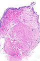
UBA - very low mag. (WC)
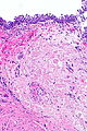
UBA - low mag. (WC)

UBA - intermed. mag. (WC)
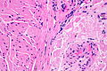
UBA - high mag. (WC)


Stains
- Congo red +ve.
Sign out
Urinary Bladder, Transurethral Resection:
- Urothelial mucosa with amyloidosis and mild chronic inflammation.
- Benign muscularis propria present.
- NEGATIVE for urothelial carcinoma in situ.
- NEGATIVE for evidence of malignancy.

Comment:
Congo red staining and polarization confirm the presence of amyloid.

Primary bladder amyloidosis is rare; systemic causes of amyloidosis should
be considered.
See also
References
- ↑ 1.0 1.1 1.2 1.3 Kobayashi, T.; Roberts, J.; Levine, J.; Degrado, J. (2014). "Primary bladder amyloidosis.". Intern Med 53 (21): 2511-3. PMID 25366012.
- ↑ 2.0 2.1 Schou-Jensen, KS.; Dahl, C.; Pilt, AP.; Azawi, NH. (Oct 2014). "Amyloidosis in the bladder: three cases with different appearance.". Scand J Urol 48 (5): 489-92. doi:10.3109/21681805.2014.920414. PMID 24857645.