Difference between revisions of "Urinary bladder amyloidosis"
Jump to navigation
Jump to search
| (4 intermediate revisions by the same user not shown) | |||
| Line 1: | Line 1: | ||
{{ Infobox diagnosis | |||
| Name = {{PAGENAME}} | |||
| Image = Urinary bladder amyloidosis -- low mag.jpg | |||
| Width = | |||
| Caption = Urinary bladder amyloidosis. [[H&E stain]]. | |||
| Synonyms = amyloidosis of the urinary bladder | |||
| Micro = lamina propria with amyloid (amorphous, paucicellular, pink material on H&E - classically "cracked") | |||
| Subtypes = | |||
| LMDDx = fibrosis, fibrin, [[leiomyoma]] | |||
| Stains = [[Congo red]] +ve | |||
| IHC = | |||
| EM = | |||
| Molecular = | |||
| IF = | |||
| Gross = | |||
| Grossing = | |||
| Site = [[urinary bladder]] | |||
| Assdx = | |||
| Syndromes = | |||
| Clinicalhx = | |||
| Signs = | |||
| Symptoms = | |||
| Prevalence = very rare | |||
| Bloodwork = | |||
| Rads = thickened bladder wall | |||
| Endoscopy = | |||
| Prognosis = | |||
| Other = | |||
| ClinDDx = other urinary bladder tumours - esp. malignant ones | |||
| Tx = | |||
}} | |||
'''Urinary bladder amyloidosis''', more formally '''primary urinary bladder amyloidosis''', is a rare benign condition of [[urinary bladder]] that can mimic [[cancer]] of the [[urinary bladder]].<ref name=pmid25366012>{{Cite journal | last1 = Kobayashi | first1 = T. | last2 = Roberts | first2 = J. | last3 = Levine | first3 = J. | last4 = Degrado | first4 = J. | title = Primary bladder amyloidosis. | journal = Intern Med | volume = 53 | issue = 21 | pages = 2511-3 | month = | year = 2014 | doi = | PMID = 25366012 }}</ref> | '''Urinary bladder amyloidosis''', more formally '''primary urinary bladder amyloidosis''', is a rare benign condition of [[urinary bladder]] that can mimic [[cancer]] of the [[urinary bladder]].<ref name=pmid25366012>{{Cite journal | last1 = Kobayashi | first1 = T. | last2 = Roberts | first2 = J. | last3 = Levine | first3 = J. | last4 = Degrado | first4 = J. | title = Primary bladder amyloidosis. | journal = Intern Med | volume = 53 | issue = 21 | pages = 2511-3 | month = | year = 2014 | doi = | PMID = 25366012 }}</ref> | ||
| Line 4: | Line 35: | ||
*Primary bladder amyloidosis is rare - approximately 200 reported cases as of 2014.<ref name=pmid25366012/><ref name=pmid24857645/> | *Primary bladder amyloidosis is rare - approximately 200 reported cases as of 2014.<ref name=pmid25366012/><ref name=pmid24857645/> | ||
**Systemic [[amyloidosis]] should be excluded.<ref name=pmid24857645>{{Cite journal | last1 = Schou-Jensen | first1 = KS. | last2 = Dahl | first2 = C. | last3 = Pilt | first3 = AP. | last4 = Azawi | first4 = NH. | title = Amyloidosis in the bladder: three cases with different appearance. | journal = Scand J Urol | volume = 48 | issue = 5 | pages = 489-92 | month = Oct | year = 2014 | doi = 10.3109/21681805.2014.920414 | PMID = 24857645 }}</ref> | **Systemic [[amyloidosis]] should be excluded.<ref name=pmid24857645>{{Cite journal | last1 = Schou-Jensen | first1 = KS. | last2 = Dahl | first2 = C. | last3 = Pilt | first3 = AP. | last4 = Azawi | first4 = NH. | title = Amyloidosis in the bladder: three cases with different appearance. | journal = Scand J Urol | volume = 48 | issue = 5 | pages = 489-92 | month = Oct | year = 2014 | doi = 10.3109/21681805.2014.920414 | PMID = 24857645 }}</ref> | ||
*May clinically mimic bladder cancer.<ref name=pmid25366012/> | *May clinically mimic [[bladder cancer]].<ref name=pmid25366012/> | ||
==Gross== | ==Gross== | ||
| Line 11: | Line 42: | ||
==Microscopic== | ==Microscopic== | ||
Features: | Features: | ||
*Lamina propria with amyloid | *Lamina propria with amyloid. | ||
**Amyloid = amorphous, paucicellular material that is [[pink on H&E]], classically has "cracked" appearance. | |||
***"Cracked": irregular fragments where the edges and centre of fragments are homogeneous. | |||
DDx: | |||
*Fibrin. | |||
*Fibrosis. | |||
*[[Leiomyoma]]. | |||
===Images=== | |||
<gallery> | |||
Image: Urinary bladder amyloidosis -- very low mag.jpg | UBA - very low mag. (WC) | |||
Image: Urinary bladder amyloidosis -- low mag.jpg | UBA - low mag. (WC) | |||
Image: Urinary bladder amyloidosis -- intermed mag.jpg | UBA - intermed. mag. (WC) | |||
Image: Urinary bladder amyloidosis -- high mag.jpg | UBA - high mag. (WC) | |||
</gallery> | |||
==Stains== | ==Stains== | ||
| Line 22: | Line 68: | ||
- Benign muscularis propria present. | - Benign muscularis propria present. | ||
- NEGATIVE for urothelial carcinoma in situ. | - NEGATIVE for urothelial carcinoma in situ. | ||
- NEGATIVE for malignancy. | - NEGATIVE for evidence of malignancy. | ||
Comment: | Comment: | ||
Congo red staining and polarization confirm the presence of amyloid. | Congo red staining and polarization confirm the presence of amyloid. | ||
Primary bladder amyloidosis is rare; systemic causes of amyloidosis should | |||
be considered. | |||
</pre> | </pre> | ||
| Line 36: | Line 83: | ||
==References== | ==References== | ||
{{Reflist|2}} | {{Reflist|2}} | ||
[[Category:Diagnosis]] | [[Category:Diagnosis]] | ||
[[Category:Genitourinary pathology]] | [[Category:Genitourinary pathology]] | ||
Latest revision as of 03:44, 23 October 2015
| Urinary bladder amyloidosis | |
|---|---|
| Diagnosis in short | |
 Urinary bladder amyloidosis. H&E stain. | |
|
| |
| Synonyms | amyloidosis of the urinary bladder |
|
| |
| LM | lamina propria with amyloid (amorphous, paucicellular, pink material on H&E - classically "cracked") |
| LM DDx | fibrosis, fibrin, leiomyoma |
| Stains | Congo red +ve |
| Site | urinary bladder |
|
| |
| Prevalence | very rare |
| Radiology | thickened bladder wall |
| Clin. DDx | other urinary bladder tumours - esp. malignant ones |
Urinary bladder amyloidosis, more formally primary urinary bladder amyloidosis, is a rare benign condition of urinary bladder that can mimic cancer of the urinary bladder.[1]
General
- Primary bladder amyloidosis is rare - approximately 200 reported cases as of 2014.[1][2]
- Systemic amyloidosis should be excluded.[2]
- May clinically mimic bladder cancer.[1]
Gross
- Urinary bladder wall thickening.[1]
Microscopic
Features:
- Lamina propria with amyloid.
- Amyloid = amorphous, paucicellular material that is pink on H&E, classically has "cracked" appearance.
- "Cracked": irregular fragments where the edges and centre of fragments are homogeneous.
- Amyloid = amorphous, paucicellular material that is pink on H&E, classically has "cracked" appearance.
DDx:
- Fibrin.
- Fibrosis.
- Leiomyoma.
Images
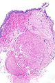
UBA - very low mag. (WC)
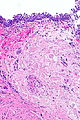
UBA - low mag. (WC)

UBA - intermed. mag. (WC)
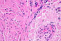
UBA - high mag. (WC)


Stains
- Congo red +ve.
Sign out
Urinary Bladder, Transurethral Resection:
- Urothelial mucosa with amyloidosis and mild chronic inflammation.
- Benign muscularis propria present.
- NEGATIVE for urothelial carcinoma in situ.
- NEGATIVE for evidence of malignancy.

Comment:
Congo red staining and polarization confirm the presence of amyloid.

Primary bladder amyloidosis is rare; systemic causes of amyloidosis should
be considered.
See also
References
- ↑ 1.0 1.1 1.2 1.3 Kobayashi, T.; Roberts, J.; Levine, J.; Degrado, J. (2014). "Primary bladder amyloidosis.". Intern Med 53 (21): 2511-3. PMID 25366012.
- ↑ 2.0 2.1 Schou-Jensen, KS.; Dahl, C.; Pilt, AP.; Azawi, NH. (Oct 2014). "Amyloidosis in the bladder: three cases with different appearance.". Scand J Urol 48 (5): 489-92. doi:10.3109/21681805.2014.920414. PMID 24857645.