Difference between revisions of "Typical carcinoid lung tumour"
Jump to navigation
Jump to search
(split out) |
(→IHC: +SO) |
||
| Line 35: | Line 35: | ||
==IHC== | ==IHC== | ||
*MIB1 scant staining. | *MIB1 scant staining. | ||
==Sign out== | |||
<pre> | |||
A. Lymph Node, Station 2L, Lymphadenectomy: | |||
- Lymph node, NEGATIVE for malignancy. | |||
B. Lymph Node, Station 4L, Lymphadenectomy: | |||
- Lymph node, NEGATIVE for malignancy. | |||
C. Lymph Node, Station 11L, Lymphadenectomy: | |||
- Lymph node, NEGATIVE for malignancy. | |||
D. Lung, Left Upper Lobe, Lobectomy: | |||
- Typical carcinoid tumour. | |||
- Carcinoid tumourlet. | |||
- Margins clear of tumour. | |||
- Please see tumour summary. | |||
</pre> | |||
==See also== | ==See also== | ||
Revision as of 13:28, 19 August 2015
Typical carcinoid lung tumour, also lung carcinoid and carcinoid tumour of the lung, is a benign lung tumour, that is excised to exclude malignancy.
General
- Approximately 80% of lung carcinoids.[1]
Presentation:[2]
- Cough.
- Hemoptysis.
Gross
- Well-circumscribed, solid.
- Location - central airways (85%), remainder peripheral.[3]
Microscopic
Features:
- Nests of cells.
- Stippled chromatin.
- Moderate cytoplasm.
- No necrosis.
- Low mitotic rate.
- Size criterion: >= 5 mm.[4][5]
DDx:
Images
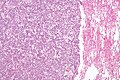
Lung carcinoid - low mag. (WC)
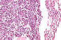
Lung carcinoid - high mag. (WC)
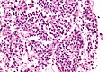
Lung carcinoid - very high mag. (WC)


IHC
- MIB1 scant staining.
Sign out
A. Lymph Node, Station 2L, Lymphadenectomy:
- Lymph node, NEGATIVE for malignancy.

B. Lymph Node, Station 4L, Lymphadenectomy:
- Lymph node, NEGATIVE for malignancy.

C. Lymph Node, Station 11L, Lymphadenectomy:
- Lymph node, NEGATIVE for malignancy.

D. Lung, Left Upper Lobe, Lobectomy:
- Typical carcinoid tumour.
- Carcinoid tumourlet.
- Margins clear of tumour.
- Please see tumour summary.
See also
References
- ↑ Naalsund, A.; Rostad, H.; Strøm, EH.; Lund, MB.; Strand, TE. (Apr 2011). "Carcinoid lung tumors--incidence, treatment and outcomes: a population-based study.". Eur J Cardiothorac Surg 39 (4): 565-9. doi:10.1016/j.ejcts.2010.08.036. PMID 20888248.
- ↑ Gungor, S.; Damadoglu, E.; Aybatli, A.; Yilmaz, A.; Kir, A.; Akkaya, E. (Jul 2006). "Typical pulmonary carcinoid tumors: presentation and outcome of 24 cases.". Med Sci Monit 12 (7): CR315-8. PMID 16810137.
- ↑ Meisinger, QC.; Klein, JS.; Butnor, KJ.; Gentchos, G.; Leavitt, BJ. (Nov 2011). "CT features of peripheral pulmonary carcinoid tumors.". AJR Am J Roentgenol 197 (5): 1073-80. doi:10.2214/AJR.10.5954. PMID 22021498.
- ↑ URL: http://pathhsw5m54.ucsf.edu/case7/image75.html. Accessed on: 23 January 2012.
- ↑ He, P.; Gu, X.; Wu, Q.; Lin, Y.; Gu, Y.; He, J. (Dec 2012). "Pulmonary carcinoid tumorlet without underlying lung disease: analysis of its relationship to fibrosis.". J Thorac Dis 4 (6): 655-8. doi:10.3978/j.issn.2072-1439.2012.06.11. PMID 23205296.
- ↑ Demirci, I.; Herold, S.; Kopp, A.; Flaßhove, M.; Klosterhalfen, B.; Janßen, H. (2012). "Overdiagnosis of a typical carcinoid tumor as an adenocarcinoma of the lung: a case report and review of the literature.". World J Surg Oncol 10: 19. doi:10.1186/1477-7819-10-19. PMID 22269186.