Difference between revisions of "Transmembrane mucin 1"
Jump to navigation
Jump to search
Jensflorian (talk | contribs) (images + refs) |
|||
| (4 intermediate revisions by the same user not shown) | |||
| Line 1: | Line 1: | ||
'''Transmembrane mucin 1''', commonly abbreviated '''MUC1''', is a commonly used [[immunostain]]. | {{ Infobox stain | ||
| Name = {{PAGENAME}} | |||
| Image = EMA_dots_ependymoma.jpg | |||
| Width = | |||
| Caption = EMA staining in ependymoma. | |||
| Abbrev = MUC1, MUC-1, EMA | |||
| Synonyms = epithelial membrane antigen | |||
| Similar = pankeratin | |||
| Clones = | |||
| Use = | |||
| Subspecial = | |||
| Pattern = | |||
| Positive = most [[adenocarcinoma]]s | |||
| Negative = [[basal cell carcinoma]] | |||
| Other = | |||
}} | |||
'''Transmembrane mucin 1''', commonly abbreviated '''MUC1''' (or '''MUC-1'''), is a commonly used [[immunostain]]. | |||
It is also known '''epithelial membrane antigen''', abbreviated '''EMA'''.<ref>{{OMIM|158340}}</ref> | It is also known '''epithelial membrane antigen''', abbreviated '''EMA'''.<ref>{{OMIM|158340}}</ref> | ||
A ring or dot-like expression pattern is typical for ependymoma. <ref>{{Cite journal | last1 = Hasselblatt | first1 = M. | last2 = Paulus | first2 = W. | title = Sensitivity and specificity of epithelial membrane antigen staining patterns in ependymomas. | journal = Acta Neuropathol | volume = 106 | issue = 4 | pages = 385-8 | month = Oct | year = 2003 | doi = 10.1007/s00401-003-0752-8 | PMID = 12898159 }}</ref> | A ring or dot-like expression pattern is typical for [[ependymoma]].<ref>{{Cite journal | last1 = Hasselblatt | first1 = M. | last2 = Paulus | first2 = W. | title = Sensitivity and specificity of epithelial membrane antigen staining patterns in ependymomas. | journal = Acta Neuropathol | volume = 106 | issue = 4 | pages = 385-8 | month = Oct | year = 2003 | doi = 10.1007/s00401-003-0752-8 | PMID = 12898159 }}</ref> | ||
===Images=== | |||
<gallery> | <gallery> | ||
File:Miningioma (4) EMA.JPG | EMA staining in a meningioma (WC/KGH) | File:Miningioma (4) EMA.JPG | EMA staining in a [[meningioma]]. (WC/KGH) | ||
File:EMA_dots_ependymoma.jpg | EMA staining in a ependymoma (WC/jensflorian) | File:EMA_dots_ependymoma.jpg | EMA staining in a ependymoma. (WC/jensflorian) | ||
</gallery> | </gallery> | ||
==See also== | ==See also== | ||
*[[Immunohistochemistry]]. | *[[Immunohistochemistry]]. | ||
*[[AE1/AE3]]. | |||
*[[Z622]]. | |||
==References== | ==References== | ||
{{Reflist|1}} | {{Reflist|1}} | ||
==External links== | |||
*[http://www.nordiqc.org/Epitopes/EMA/EMA.htm EMA (nordiqc.org)]. | |||
[[Category:Immunohistochemistry]] | [[Category:Immunohistochemistry]] | ||
Latest revision as of 19:52, 4 January 2019
| Transmembrane mucin 1 | |
|---|---|
| Stain in short | |
 EMA staining in ependymoma. | |
| Abbreviation | MUC1, MUC-1, EMA |
| Synonyms | epithelial membrane antigen |
| Similar stains | pankeratin |
| Positive | most adenocarcinomas |
| Negative | basal cell carcinoma |
Transmembrane mucin 1, commonly abbreviated MUC1 (or MUC-1), is a commonly used immunostain.
It is also known epithelial membrane antigen, abbreviated EMA.[1]
A ring or dot-like expression pattern is typical for ependymoma.[2]
Images
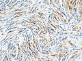
EMA staining in a meningioma. (WC/KGH)
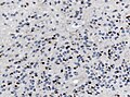
EMA staining in a ependymoma. (WC/jensflorian)
_EMA.JPG)
See also
References
- ↑ Online 'Mendelian Inheritance in Man' (OMIM) 158340
- ↑ Hasselblatt, M.; Paulus, W. (Oct 2003). "Sensitivity and specificity of epithelial membrane antigen staining patterns in ependymomas.". Acta Neuropathol 106 (4): 385-8. doi:10.1007/s00401-003-0752-8. PMID 12898159.