Difference between revisions of "Total nephrectomy for tumour grossing"
Jump to navigation
Jump to search
| (14 intermediate revisions by the same user not shown) | |||
| Line 1: | Line 1: | ||
[[Image:Renal oncocytoma.jpg|right|thumb|A kidney tumour ([[renal oncocytoma]]) at the time of grossing.]] | |||
This article deals with the [[cut-up]] of '''total nephrectomy for tumour''' specimens. It also includes '''radical nephrectomy''' with or without an adrenal gland. | This article deals with the [[cut-up]] of '''total nephrectomy for tumour''' specimens. It also includes '''radical nephrectomy''' with or without an adrenal gland. | ||
''Partial nephrectomy'' specimens are dealt with separately. | ''[[Partial nephrectomy]]'' specimens and ''[[nephroureterectomy]]'' specimens are dealt with separately. | ||
==Introduction== | ==Introduction== | ||
| Line 10: | Line 11: | ||
*Total nephrectomy. | *Total nephrectomy. | ||
*Radical nephrectomy - includes Gerota's fascia. | *Radical nephrectomy - includes Gerota's fascia. | ||
**Gerota's fascia is the fascia overlying the perinephric fat | **Gerota's fascia is the fascia overlying the perinephric fat. | ||
**''Radical nephrectomy'' by definition does ''not'' require removal of the [[adrenal gland]].<ref name=pmid26425218>{{Cite journal | last1 = Nason | first1 = GJ. | last2 = Walsh | first2 = LG. | last3 = Redmond | first3 = CE. | last4 = Kelly | first4 = NP. | last5 = McGuire | first5 = BB. | last6 = Sharma | first6 = V. | last7 = Kelly | first7 = ME. | last8 = Galvin | first8 = DJ. | last9 = Mulvin | first9 = DW. | title = Comparative effectiveness of adrenal sparing radical nephrectomy and non-adrenal sparing radical nephrectomy in clear cell renal cell carcinoma: Observational study of survival outcomes. | journal = Can Urol Assoc J | volume = 9 | issue = 9-10 | pages = E583-8 | month = | year = | doi = 10.5489/cuaj.2842 | PMID = 26425218 }}</ref><ref name=pmid23039377>{{Cite journal | last1 = Yap | first1 = SA. | last2 = Alibhai | first2 = SM. | last3 = Abouassaly | first3 = R. | last4 = Timilshina | first4 = N. | last5 = Margel | first5 = D. | last6 = Finelli | first6 = A. | title = Ipsilateral adrenalectomy at the time of radical nephrectomy impacts overall survival. | journal = BJU Int | volume = 111 | issue = 3 Pt B | pages = E54-8 | month = Mar | year = 2013 | doi = 10.1111/j.1464-410X.2012.11435.x | PMID = 23039377 }}</ref> | |||
Resections for tumours generally are ''radical nephrectomies'' or ''partial nephrectomies''. | Resections for tumours generally are ''radical nephrectomies'' or ''partial nephrectomies''. | ||
| Line 24: | Line 26: | ||
===Opening notes=== | ===Opening notes=== | ||
*It is reasonable to skip the inking step as if the tumour is at the surface it is usually obvious. | *It is reasonable to skip the [[inking]] step as if the tumour is at the surface it is usually obvious. | ||
==Protocol== | ==Protocol== | ||
| Line 51: | Line 53: | ||
*Margin: [nearest margin ___, distance ___ cm / positive margin, location ___]. | *Margin: [nearest margin ___, distance ___ cm / positive margin, location ___]. | ||
*Extension into perinephric fat: [absent / not identified-pushing border / suspicious / present]. | *Extension into perinephric fat: [absent / not identified-pushing border / suspicious / present]. | ||
*Extension into renal | *Extension into renal sinus fat: [absent / not identified-pushing border / suspicious / present]. | ||
*Extension into the collecting system: [absent / suspicious / present]. | *Extension into the collecting system: [absent / suspicious / present]. | ||
*Extension into renal vein: [absent / suspicious / present]. | *Extension into renal vein: [absent / suspicious / present]. | ||
| Line 65: | Line 67: | ||
*Tumour with nearest margin. | *Tumour with nearest margin. | ||
*Tumour and perinephric fat. † | *Tumour and perinephric fat. † | ||
*Tumour and | *Tumour and sinus fat. † | ||
*Normal kidney. | *Normal kidney. | ||
*Adrenal gland. | *Adrenal gland. | ||
===Protocol notes=== | ===Protocol notes=== | ||
* | *[[Kidney cancer staging|Kidney tumour stage]] size cut points: <=4 cm, <=7 cm. | ||
*‡ It is important to sample the renal vein wall if tumour thrombus projecting out of the renal vein, as a positive margin is called based on microscopic involvement of | **The 7th edition of the [[TNM staging system]] divides pT2 into pT2a (>7 cm and <=10 cm) and pT2b (>10 cm); however, evidence does not support this subdivision.<ref name=pmid21030143>{{Cite journal | last1 = Waalkes | first1 = S. | last2 = Becker | first2 = F. | last3 = Schrader | first3 = AJ. | last4 = Janssen | first4 = M. | last5 = Wegener | first5 = G. | last6 = Merseburger | first6 = AS. | last7 = Schrader | first7 = M. | last8 = Hofmann | first8 = R. | last9 = Stöckle | first9 = M. | title = Is there a need to further subclassify pT2 renal cell cancers as implemented by the revised 7th TNM version? | journal = Eur Urol | volume = 59 | issue = 2 | pages = 258-63 | month = Feb | year = 2011 | doi = 10.1016/j.eururo.2010.10.005 | PMID = 21030143 }}</ref> | ||
*‡ It is important to sample the renal vein wall if tumour thrombus is projecting out of the renal vein, as a positive margin is called based on microscopic involvement of the vein or tumour adherence to the vein wall at microscopy.<ref name=pmid24025521>{{Cite journal | last1 = Trpkov | first1 = K. | last2 = Grignon | first2 = DJ. | last3 = Bonsib | first3 = SM. | last4 = Amin | first4 = MB. | last5 = Billis | first5 = A. | last6 = Lopez-Beltran | first6 = A. | last7 = Samaratunga | first7 = H. | last8 = Tamboli | first8 = P. | last9 = Delahunt | first9 = B. | title = Handling and staging of renal cell carcinoma: the International Society of Urological Pathology Consensus (ISUP) conference recommendations. | journal = Am J Surg Pathol | volume = 37 | issue = 10 | pages = 1505-17 | month = Oct | year = 2013 | doi = 10.1097/PAS.0b013e31829a85d0 | PMID = 24025521 }}</ref> | |||
**Tumour projecting out of the vein (i.e. at the surface of specimen), at the time of grossing, is presumed to be due to retraction of the vein after it is cut. | **Tumour projecting out of the vein (i.e. at the surface of specimen), at the time of grossing, is presumed to be due to retraction of the vein after it is cut. | ||
*† If fat invasion obvious = 1 section. | *† If fat invasion obvious = 1 section. | ||
**Suspicion of fat invasion = 3 sections. | **Suspicion of fat invasion = 3 sections. | ||
***It should be noted that as per Trpkov ''et al.''<ref name=pmid24025521/>: "<nowiki>[A]</nowiki> circumscribed, pushing ... border, even if extending well beyond the normal outline of the renal cortex, is '''not''' <nowiki>[emphasis added]</nowiki> diagnostic of perinephric fat invasion." | |||
===Alternate approaches=== | ===Alternate approaches=== | ||
*Ink only the areas that are suspicious for tumour. | |||
==See also== | ==See also== | ||
*[[Kidney cancer staging]]. | |||
*[[Marking ink]]. | |||
===Related protocols=== | ===Related protocols=== | ||
*[[Partial nephrectomy]]. | *[[Partial nephrectomy]]. | ||
*[[Nephroureterectomy]]. | |||
==References== | ==References== | ||
{{Reflist|1}} | {{Reflist|1}} | ||
Latest revision as of 16:21, 6 July 2017

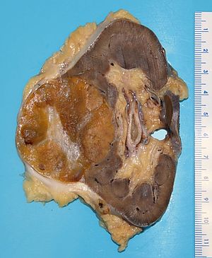
A kidney tumour (renal oncocytoma) at the time of grossing.
This article deals with the cut-up of total nephrectomy for tumour specimens. It also includes radical nephrectomy with or without an adrenal gland.
Partial nephrectomy specimens and nephroureterectomy specimens are dealt with separately.
Introduction
Nephrectomies are often done for kidney tumours.
They come in three basics flavours:
- Partial nephrectomy.
- Total nephrectomy.
- Radical nephrectomy - includes Gerota's fascia.
- Gerota's fascia is the fascia overlying the perinephric fat.
- Radical nephrectomy by definition does not require removal of the adrenal gland.[1][2]
Resections for tumours generally are radical nephrectomies or partial nephrectomies.
Specimen opening
- Paint surface of specimen (optional).
- Take margins - renal artery, renal vein, ureter (all en face) and place all three in one tissue cassette.
- Place probes in the renal vein.
- One toward upper pole.
- One toward lower pole.
- Cut the kidney in the frontal plane (from medial to lateral) using the two probes as a guide.
- The cut should open the renal vein and its major tributaries.
Opening notes
- It is reasonable to skip the inking step as if the tumour is at the surface it is usually obvious.
Protocol
Dimensions, weight and inking:
- Type: [total nephrectomy/radical nephrectomy].
- Laterality: [left / right].
- Weight: ___ grams.
- Size of specimen (superior-inferior, left-right, anterior-posterior): ___ x ___ x ___ cm.
- Ureter (length x diameter): ___ x ___ cm.
- Renal vein (length x diameter): ___ x ___ cm.
- Renal artery (length x diameter): ___ x ___ cm.
- Adrenal gland: [___ x ___ x ___ cm / not identified].
- Inking of surface: [colour].
- Size of kidney (superior-inferior, left-right, anterior-posterior): ___ x ___ x ___ cm.
- Perinephric fat (maximal dimension): ___ cm.
Tumour:
- Dimensions (superior-inferior, left-right, anterior-posterior): ___ x ___ x ___ cm.
- Location: [upper pole / mid / lower pole].
- Colour: [yellow / tan / white].
- Morphology: [solid / cystic / solid and cystic - with ___ % cystic].
- Friability: [friable / not friable].
- Circumscription: [well circumscribed / indeterminate / infiltrative border].
- Hemorrhage: [present / absent].
- Necrosis: [present / absent].
- Margin: [nearest margin ___, distance ___ cm / positive margin, location ___].
- Extension into perinephric fat: [absent / not identified-pushing border / suspicious / present].
- Extension into renal sinus fat: [absent / not identified-pushing border / suspicious / present].
- Extension into the collecting system: [absent / suspicious / present].
- Extension into renal vein: [absent / suspicious / present].
Other:
- Non-tumour parenchyma: [cortex unremarkable / thinned].
- Collecting system mucosa: [smooth and regular / granular / irregular / dilated].
- Lymph nodes: [number of lymph nodes with [unremarkable cut surface / tumour] / not identified].
Representative sections are submitted:
- Renal vein margin. ‡
- Ureter margin and renal artery margin.
- Tumour with nearest margin.
- Tumour and perinephric fat. †
- Tumour and sinus fat. †
- Normal kidney.
- Adrenal gland.
Protocol notes
- Kidney tumour stage size cut points: <=4 cm, <=7 cm.
- The 7th edition of the TNM staging system divides pT2 into pT2a (>7 cm and <=10 cm) and pT2b (>10 cm); however, evidence does not support this subdivision.[3]
- ‡ It is important to sample the renal vein wall if tumour thrombus is projecting out of the renal vein, as a positive margin is called based on microscopic involvement of the vein or tumour adherence to the vein wall at microscopy.[4]
- Tumour projecting out of the vein (i.e. at the surface of specimen), at the time of grossing, is presumed to be due to retraction of the vein after it is cut.
- † If fat invasion obvious = 1 section.
- Suspicion of fat invasion = 3 sections.
- It should be noted that as per Trpkov et al.[4]: "[A] circumscribed, pushing ... border, even if extending well beyond the normal outline of the renal cortex, is not [emphasis added] diagnostic of perinephric fat invasion."
- Suspicion of fat invasion = 3 sections.
Alternate approaches
- Ink only the areas that are suspicious for tumour.
See also
Related protocols
References
- ↑ Nason, GJ.; Walsh, LG.; Redmond, CE.; Kelly, NP.; McGuire, BB.; Sharma, V.; Kelly, ME.; Galvin, DJ. et al. "Comparative effectiveness of adrenal sparing radical nephrectomy and non-adrenal sparing radical nephrectomy in clear cell renal cell carcinoma: Observational study of survival outcomes.". Can Urol Assoc J 9 (9-10): E583-8. doi:10.5489/cuaj.2842. PMID 26425218.
- ↑ Yap, SA.; Alibhai, SM.; Abouassaly, R.; Timilshina, N.; Margel, D.; Finelli, A. (Mar 2013). "Ipsilateral adrenalectomy at the time of radical nephrectomy impacts overall survival.". BJU Int 111 (3 Pt B): E54-8. doi:10.1111/j.1464-410X.2012.11435.x. PMID 23039377.
- ↑ Waalkes, S.; Becker, F.; Schrader, AJ.; Janssen, M.; Wegener, G.; Merseburger, AS.; Schrader, M.; Hofmann, R. et al. (Feb 2011). "Is there a need to further subclassify pT2 renal cell cancers as implemented by the revised 7th TNM version?". Eur Urol 59 (2): 258-63. doi:10.1016/j.eururo.2010.10.005. PMID 21030143.
- ↑ 4.0 4.1 Trpkov, K.; Grignon, DJ.; Bonsib, SM.; Amin, MB.; Billis, A.; Lopez-Beltran, A.; Samaratunga, H.; Tamboli, P. et al. (Oct 2013). "Handling and staging of renal cell carcinoma: the International Society of Urological Pathology Consensus (ISUP) conference recommendations.". Am J Surg Pathol 37 (10): 1505-17. doi:10.1097/PAS.0b013e31829a85d0. PMID 24025521.