Subependymoma
Jump to navigation
Jump to search
| Subependymoma | |
|---|---|
| Diagnosis in short | |
 Subependymoma. H&E stain. | |
|
| |
| LM | microcysts with bluish material (give a spongy appearance at low magnification), clustering of nuclei cluster (described as "bundles of flowers"), bland nuclei |
| Site | brain - see neuropathology tumours |
|
| |
| Symptoms | +/-headache |
| Radiology | classically fourth ventricle |
| Prognosis | WHO grade I |
| Clin. DDx | other brain tumours - ependymoma, CNS lymphoma |
| Treatment | surgical excision |
Subependymoma is neuropathology tumour classically found in the fourth ventricle.
General
- Good prognosis - WHO Grade I.
- Low-grade glial tumour.[1]
Clinical:[1]
- Slow growing.
- +/-Headaches.
- Tx: surgery.
Gross/radiology
- Classic location: fourth ventricle.[2]
- Well-demarcated margin.
- Usu. completely within the ventricle; does not extend into brain (like ependymomas).
Microscopic
Features:[3]
- Microcysts with bluish material - give a spongy appearance at low magnification.
- Nuclei cluster.
- Described as "bundles of flowers".
Negatives.
- No nuclear pleomorphism, no prominent nucleoli, no mitoses.
- Do not invade into brain.[1]
Images
www:
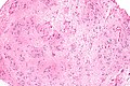
Subependyoma - intermed. mag. (WC)
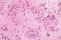
Subependymoma - high mag. (WC)
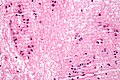
Subependymoma - very high mag. (WC)


See also
References
- ↑ 1.0 1.1 1.2 Castro-Castro, J.; Castro-Bouzas, D.; Prieto-Casal, PL.; Carcacia-Hermilla, ID.; Riu-Lloveras, M.; Castro-Gómez, JE. (Mar 2013). "[Subependymoma of the lateral ventricle. A case report].". Rev Neurol 56 (6): 332-6. PMID 23483468.
- ↑ Hoeffel, C.; Boukobza, M.; Polivka, M.; Lot, G.; Guichard, JP.; Lafitte, F.; Reizine, D.; Merland, JJ.. "MR manifestations of subependymomas.". AJNR Am J Neuroradiol 16 (10): 2121-9. PMID 8585504. http://www.ajnr.org/cgi/reprint/16/10/2121.
- ↑ 3.0 3.1 URL: http://moon.ouhsc.edu/kfung/jty1/Com05/Com501-2-Diss.htm. Accessed on: 2 June 2011.