Difference between revisions of "Renal rhabdoid tumour"
Jump to navigation
Jump to search
(split out) |
(+image) |
||
| Line 14: | Line 14: | ||
*Prominent [[nucleolus]] -- '''key feature'''. | *Prominent [[nucleolus]] -- '''key feature'''. | ||
Images: | ===Images=== | ||
<gallery> | |||
Image:Rhabdoid tumor.jpg | Rhabdoid tumour. (WC/AFIP) | |||
</gallery> | |||
www: | |||
*[http://www.flickr.com/photos/ckrishnan/3954115280/in/photostream RTK - low mag. (flickr.com)]. | *[http://www.flickr.com/photos/ckrishnan/3954115280/in/photostream RTK - low mag. (flickr.com)]. | ||
*[http://www.flickr.com/photos/ckrishnan/3953336593/in/photostream RTK - high mag. (flickr.com)]. | *[http://www.flickr.com/photos/ckrishnan/3953336593/in/photostream RTK - high mag. (flickr.com)]. | ||
Revision as of 03:07, 4 April 2015
Renal rhabdoid tumour is a rare malignant tumour of the kidney seen in children.
General
- Similar to extrarenal malignant rhabdoid tumour.[1]
- Arises from renal medulla.
- May be associated with a CNS tumour.
Microscopic
- Variable architecture.
- Round cells.
- Abundant cytoplasm with eosinophilic inclusions.
- Eccentric vesicular nucleus.
- Prominent nucleolus -- key feature.
Images
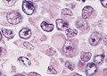
Rhabdoid tumour. (WC/AFIP)

www:
IHC
- INI1 -ve.
See also
References
- ↑ 1.0 1.1 Humphrey, Peter A; Dehner, Louis P; Pfeifer, John D (2008). The Washington Manual of Surgical Pathology (1st ed.). Lippincott Williams & Wilkins. pp. 284. ISBN 978-0781765275.
- ↑ Humphrey, Peter A; Dehner, Louis P; Pfeifer, John D (2008). The Washington Manual of Surgical Pathology (1st ed.). Lippincott Williams & Wilkins. pp. 627. ISBN 978-0781765275.