Difference between revisions of "Renal oncocytoma"
Jump to navigation
Jump to search
(+cat.) |
(split-out) |
||
| Line 1: | Line 1: | ||
'''Renal oncocytoma''' is a benign [[kidney tumour]] that is removed to exclude malignancy. | |||
==General== | |||
*Can be difficult to distinguish radiologically from RCC (chromophobe subtype). | |||
** ... and pathologists occasionally struggle like the radiologists. | |||
*Benign tumour - the reason it is excised is... one cannot be certain it isn't a RCC. | |||
==Gross== | |||
*Brown, mahogany brown. | |||
*1/3 have a characteristic central scar.<ref name=Ref_GUP302>{{Ref GUP|302}}</ref> | |||
===Image=== | |||
<gallery> | |||
Image:Renal_oncocytoma.jpg| Renal oncocytoma with central scar. (WP) | |||
</gallery> | |||
==Microscopic== | |||
Features: | |||
*Eosinophilic cytoplasm - slightly granular '''key feature'''. | |||
*Cells arranged in nests. | |||
*Nuclei uniform and round.<ref name=Ref_GUP302>{{Ref GUP|302}}</ref> | |||
**Slightly enlarged nuclei, but '''no significant''' pleomorphism (size variation) - '''important'''. | |||
Notes: | |||
*May look like eosinophilic variant of chromophobe RCC -- this is the main DDx. | |||
**A comparison based on histomorphology: ''[[Kidney_tumours#Tabular_comparison_of_oncocytoma_and_chromophobe_RCC|Tabular comparison between ChRCC & oncocytoma]]''. | |||
***Oncocytoma typically has: no perinuclear clearing, no raisinoid nuclei, no binucleation. | |||
DDx: | |||
*[[Chromophobe renal cell carcinoma]], eosinophilic variant. | |||
*[[Clear cell renal cell carcinoma]], eosinophilic variant. | |||
===Images=== | |||
<gallery> | |||
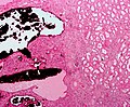
Image:Renal_oncocytoma2.jpg | Oncocytoma - high mag. (WC/Nephron) | |||
Image:Renal_oncocytoma3.jpg | Oncocytoma - intermed. mag. (WC/Nephron) | |||
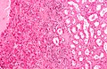
Image:Renal_oncocytoma4.jpg | Oncocytoma - low mag. (WC/Nephron) | |||
</gallery> | |||
==Stain=== | |||
*[[Hale's colloidal iron]] -ve | |||
**[[ChRCC]] +ve (blue granular cytoplasmic). | |||
===IHC=== | |||
*CK7 -ve<ref name=pmid17683191>{{Cite journal | last1 = Liu | first1 = L. | last2 = Qian | first2 = J. | last3 = Singh | first3 = H. | last4 = Meiers | first4 = I. | last5 = Zhou | first5 = X. | last6 = Bostwick | first6 = DG. | title = Immunohistochemical analysis of chromophobe renal cell carcinoma, renal oncocytoma, and clear cell carcinoma: an optimal and practical panel for differential diagnosis. | journal = Arch Pathol Lab Med | volume = 131 | issue = 8 | pages = 1290-7 | month = Aug | year = 2007 | doi = 10.1043/1543-2165(2007)131[1290:IAOCRC]2.0.CO;2 | PMID = 17683191 }}</ref>/+ve (cytoplasmic) . | |||
**Chromophobe renal cell carcinoma = cell membrane +ve. | |||
==Sign out== | |||
<pre> | |||
PORTION OF KIDNEY, RIGHT, PARTIAL NEPHRECTOMY: | |||
- ONCOCYTOMA. | |||
</pre> | |||
===Micro=== | |||
The sections show a tumour with cells arranged in nests. The tumour cells have abundant | |||
eosinophilic cytoplasm. The tumour cell nuclei are round and have round nucleoli. No | |||
perinuclear halos are apparent. Binucleation is not apparent. No zonal necrosis is | |||
identified. Focally, tumour nests are spaced reminiscent of an archipelago. Mitoses are not | |||
apparent. The tumour is moderately circumscribed. | |||
The thin rim of renal parenchyma has no apparent pathology on the H&E stained sections. | |||
==See also== | |||
*[[Kidney tumours]]. | |||
*[[Oncocytoma]]. | |||
==References== | |||
{{Reflist|2}} | |||
[[Category:Kidney tumours]] | |||
[[Category:Diagnosis]] | [[Category:Diagnosis]] | ||
Revision as of 03:52, 4 November 2013
Renal oncocytoma is a benign kidney tumour that is removed to exclude malignancy.
General
- Can be difficult to distinguish radiologically from RCC (chromophobe subtype).
- ... and pathologists occasionally struggle like the radiologists.
- Benign tumour - the reason it is excised is... one cannot be certain it isn't a RCC.
Gross
- Brown, mahogany brown.
- 1/3 have a characteristic central scar.[1]
Image
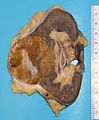
Renal oncocytoma with central scar. (WP)

Microscopic
Features:
- Eosinophilic cytoplasm - slightly granular key feature.
- Cells arranged in nests.
- Nuclei uniform and round.[1]
- Slightly enlarged nuclei, but no significant pleomorphism (size variation) - important.
Notes:
- May look like eosinophilic variant of chromophobe RCC -- this is the main DDx.
- A comparison based on histomorphology: Tabular comparison between ChRCC & oncocytoma.
- Oncocytoma typically has: no perinuclear clearing, no raisinoid nuclei, no binucleation.
- A comparison based on histomorphology: Tabular comparison between ChRCC & oncocytoma.
DDx:
- Chromophobe renal cell carcinoma, eosinophilic variant.
- Clear cell renal cell carcinoma, eosinophilic variant.
Images
Oncocytoma - high mag. (WC/Nephron)
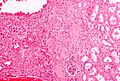
Oncocytoma - intermed. mag. (WC/Nephron)
Oncocytoma - low mag. (WC/Nephron)


Stain=
- Hale's colloidal iron -ve
- ChRCC +ve (blue granular cytoplasmic).
IHC
- CK7 -ve[2]/+ve (cytoplasmic) .
- Chromophobe renal cell carcinoma = cell membrane +ve.
Sign out
PORTION OF KIDNEY, RIGHT, PARTIAL NEPHRECTOMY:
- ONCOCYTOMA.
Micro
The sections show a tumour with cells arranged in nests. The tumour cells have abundant eosinophilic cytoplasm. The tumour cell nuclei are round and have round nucleoli. No perinuclear halos are apparent. Binucleation is not apparent. No zonal necrosis is identified. Focally, tumour nests are spaced reminiscent of an archipelago. Mitoses are not apparent. The tumour is moderately circumscribed.
The thin rim of renal parenchyma has no apparent pathology on the H&E stained sections.
See also
References
- ↑ 1.0 1.1 Zhou, Ming; Magi-Galluzzi, Cristina (2006). Genitourinary Pathology: A Volume in Foundations in Diagnostic Pathology Series (1st ed.). Churchill Livingstone. pp. 302. ISBN 978-0443066771.
- ↑ Liu, L.; Qian, J.; Singh, H.; Meiers, I.; Zhou, X.; Bostwick, DG. (Aug 2007). "Immunohistochemical analysis of chromophobe renal cell carcinoma, renal oncocytoma, and clear cell carcinoma: an optimal and practical panel for differential diagnosis.". Arch Pathol Lab Med 131 (8): 1290-7. doi:10.1043/1543-2165(2007)131[1290:IAOCRC]2.0.CO;2. PMID 17683191.