Difference between revisions of "Red blood cell"
Jump to navigation
Jump to search
(create) |
|||
| (19 intermediate revisions by the same user not shown) | |||
| Line 1: | Line 1: | ||
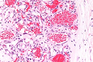
The '''red blood cell'', abbreviated '''RBC''', is the carrier of oxygen to tissue. It is seen daily by pathologists. | [[Image:Capillary hemangioma - very high mag.jpg|thumb|right|300px|Abundant red blood cells in the vascular channels of a [[hemangioma]]. [[H&E stain|H&E stain]].]] | ||
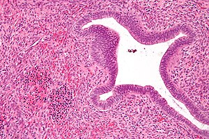
[[Image:Nucleated_red_blood_cells_-_endometrial_polyp_-_high_mag.jpg|thumb|300px|Nucleated red blood cells in an [[endometrial polyp]]. [[H&E stain]].]] | |||
The '''red blood cell''', abbreviated '''RBC''', is the carrier of oxygen to tissue. It is seen daily by pathologists. | |||
It is approximately 8 | It is approximately 8 [[micrometer]]s in diameter.<ref>URL: [http://www.wisegeek.com/how-large-is-a-micrometer.htm http://www.wisegeek.com/how-large-is-a-micrometer.htm]. Accessed on: 17 January 2011.</ref> | ||
==See also | =Precursors= | ||
===Reticulocyte=== | |||
The direct precursor to the RBC is the '''reticulocyte'''. | |||
Image: | |||
*[http://commons.wikimedia.org/wiki/File:Reticulocytes_Human_Blood_Supravital_Stain.jpg Reticulocytes (WC)]. | |||
===Normoblast=== | |||
'''Normoblasts''' are the nucleated precursors of RBCs. | |||
Images: | |||
*[http://commons.wikimedia.org/wiki/File:Hematopoiesis_%28human%29_diagram.png Hematopoiesis diagram (WC)]. | |||
*[http://commons.wikimedia.org/wiki/File:Orthochromatic_erythroblast.png Normoblast (WC)]. | |||
=Conditions with RBCs= | |||
==Sickle cell disease== | |||
{{Main|Sickle cell disease}} | |||
==Anemia== | |||
{{Main|Anemia}} | |||
==Hemophagocytic syndrome== | |||
{{Main|Hemophagocytic syndrome}} | |||
*Macrophages eat whole RBCs. | |||
==Myospherulosis== | |||
*[[AKA]] ''spherulocytosis''.<ref name=pmid7591868/> | |||
===General=== | |||
*Foreign body-type [[granuloma|granulomatous]] reaction to lipid-containing material and blood.<ref name=pmid7591868/><ref name=pmid11811513>{{Cite journal | last1 = Fisher | first1 = SC. | last2 = Horning | first2 = GM. | last3 = Hellstein | first3 = JW. | title = Myospherulosis complicating cortical block grafting: a case report. | journal = J Periodontol | volume = 72 | issue = 12 | pages = 1755-9 | month = Dec | year = 2001 | doi = 10.1902/jop.2001.72.12.1755 | PMID = 11811513 }}</ref> | |||
*Rare.<ref name=pmid9866916>{{Cite journal | last1 = Sarkar | first1 = S. | last2 = Gangane | first2 = N. | last3 = Sharma | first3 = S. | title = Myospherulosis of maxillary sinus--a case report with review of literature. | journal = Indian J Pathol Microbiol | volume = 41 | issue = 4 | pages = 491-3 | month = Oct | year = 1998 | doi = | PMID = 9866916 }}</ref> | |||
Etiology: | |||
*Exposure to dying fat,<ref name=pmid7591868>{{Cite journal | last1 = Godbersen | first1 = GS. | last2 = Kleeberg | first2 = J. | last3 = Lüttges | first3 = J. | last4 = Werner | first4 = JA. | title = [Spherulocytosis (myospherulosis) of the paranasal sinuses]. | journal = HNO | volume = 43 | issue = 9 | pages = 552-5 | month = Sep | year = 1995 | doi = | PMID = 7591868 }}</ref> e.g. [[fat necrosis of the breast]]. | |||
*Malignancy, e.g. [[renal cell carcinoma]].<ref name=pmid11035579>{{Cite journal | last1 = Chau | first1 = KY. | last2 = Pretorius | first2 = JM. | last3 = Stewart | first3 = AW. | title = Myospherulosis in renal cell carcinoma. | journal = Arch Pathol Lab Med | volume = 124 | issue = 10 | pages = 1476-9 | month = Oct | year = 2000 | doi = 10.1043/0003-9985(2000)1241476:MIRCC2.0.CO;2 | PMID = 11035579 }}</ref> | |||
===Microscopic=== | |||
Features: | |||
*Phagocytosed RBCs. | |||
**Round aggregates of red blood cells ~10-20 RBCs in diameter (80-160 micrometers). | |||
=See also= | |||
*[[Neutrophil]]. | *[[Neutrophil]]. | ||
*[[Eosinophil]]. | |||
*[[Red blood cell extravasation]]. | |||
*[[Amebiasis]]. | |||
=References= | |||
{{Reflist| | {{Reflist|2}} | ||
[[Category:Basics]] | [[Category:Basics]] | ||
Latest revision as of 11:09, 25 November 2016


The red blood cell, abbreviated RBC, is the carrier of oxygen to tissue. It is seen daily by pathologists.
It is approximately 8 micrometers in diameter.[1]
Precursors
Reticulocyte
The direct precursor to the RBC is the reticulocyte.
Image:
Normoblast
Normoblasts are the nucleated precursors of RBCs.
Images:
Conditions with RBCs
Sickle cell disease
Main article: Sickle cell disease
Anemia
Main article: Anemia
Hemophagocytic syndrome
Main article: Hemophagocytic syndrome
- Macrophages eat whole RBCs.
Myospherulosis
General
- Foreign body-type granulomatous reaction to lipid-containing material and blood.[2][3]
- Rare.[4]
Etiology:
- Exposure to dying fat,[2] e.g. fat necrosis of the breast.
- Malignancy, e.g. renal cell carcinoma.[5]
Microscopic
Features:
- Phagocytosed RBCs.
- Round aggregates of red blood cells ~10-20 RBCs in diameter (80-160 micrometers).
See also
References
- ↑ URL: http://www.wisegeek.com/how-large-is-a-micrometer.htm. Accessed on: 17 January 2011.
- ↑ 2.0 2.1 2.2 Godbersen, GS.; Kleeberg, J.; Lüttges, J.; Werner, JA. (Sep 1995). "[Spherulocytosis (myospherulosis) of the paranasal sinuses].". HNO 43 (9): 552-5. PMID 7591868.
- ↑ Fisher, SC.; Horning, GM.; Hellstein, JW. (Dec 2001). "Myospherulosis complicating cortical block grafting: a case report.". J Periodontol 72 (12): 1755-9. doi:10.1902/jop.2001.72.12.1755. PMID 11811513.
- ↑ Sarkar, S.; Gangane, N.; Sharma, S. (Oct 1998). "Myospherulosis of maxillary sinus--a case report with review of literature.". Indian J Pathol Microbiol 41 (4): 491-3. PMID 9866916.
- ↑ Chau, KY.; Pretorius, JM.; Stewart, AW. (Oct 2000). "Myospherulosis in renal cell carcinoma.". Arch Pathol Lab Med 124 (10): 1476-9. doi:10.1043/0003-9985(2000)1241476:MIRCC2.0.CO;2. PMID 11035579.