Pulmonary hamartoma
| Pulmonary hamartoma | |
|---|---|
| Diagnosis in short | |
 Pulmonary hamartoma. H&E stain. | |
|
| |
| LM | benign cartilage, adipocytes and respiratory epithelium; lesion without significant nuclear atypia |
| Gross | well circumscribed, cartilageous or fatty appearing |
| Site | lung |
|
| |
| Prevalence | uncommon |
| Prognosis | benign |
| Clin. DDx | slow growing lung tumours |
Pulmonary hamartoma, also lung hamartoma, is a benign lesion of the lung that may be confused with malignancy.
General
- Benign.
See also: Hamartoma.
Gross
- Well circumscribed lesion.
Microscopic
Features:
- Cartilage - key feature.
- Single cells in lacunae surrounded by abundant matrix.
- Paucicellular vis-a-vis malignant lesions.
- Single cells in lacunae surrounded by abundant matrix.
- Fat (adipocytes) - key feature.
- Respiratory epithelium (columnar epithelium with cilia).
Notes:
- No nuclear atypia.
DDx:
- Other lung tumours - especially slow growing ones.
Images
www:
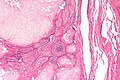
Pulmonary hamartoma - low mag. (WC)
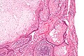
Pulmonary hamartoma - intermed. mag. (WC)
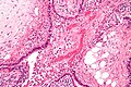
Pulmonary hamartoma - high mag. (WC)


IHC
- S100 +ve - highlights the fat.
Sign out
LUNG LESION, LEFT UPPER LOBE, WEDGE RESECTION:
- PULMONARY HAMARTOMA WITH MILD FOCAL ACUTE INFLAMMATION AND SURROUNDING EDEMA.
- SURROUNDING LUNG WITH MILD EMPHYSEMATOUS CHANGES.
Micro
The sections show lung with a well circumscribed lesion with a fibrous capsule partially lined by respiratory-type epithelium. The lesion consists of abundant respiratory epithelium and glands with focal sheeting and small collections of neutrophils focally. Small foci of degenerative changes are seen. The epithelium of the lesion as a bland cytomorphology. Mitotic activity is not readily apparent. Fat is not identified as a component of the lesion. Around the periphery of the lesion pulmonary edema is present.
The piece surrounding lung more distant from the lesion has mild emphysematous changes. No interstitial fibrosis is identified. No significant inflammation is present. The arteries are approximately the size of accompanying airway. The arteries have no appreciable intimal thickening.
See also
References
- ↑ URL: http://www.path.utah.edu/casepath/pm%20cases/pmcase8/pmcase8part4.htm. Accessed on: 9 June 2011.